5CRE | pdb_00005cre

Human skeletal calsequestrin, D210G mutant low-calcium complex


Experimental Data Snapshot

  • Method: X-RAY DIFFRACTION
  • Resolution: 3.31 Å
  • R-Value Free: 
    0.290 (Depositor), 0.289 (DCC) 
  • R-Value Work: 
    0.249 (Depositor), 0.251 (DCC) 
  • R-Value Observed: 
    0.253 (Depositor) 

Starting Model: experimental
View more details

wwPDB Validation 3D Report Full Report

Validation slider image for 5CRE

This is version 1.3 of the entry. See complete history

Literature

Characterization of Two Human Skeletal Calsequestrin Mutants Implicated in Malignant Hyperthermia and Vacuolar Aggregate Myopathy.

Lewis, K.M.Ronish, L.A.Rios, E.Kang, C.

(2015) J Biological Chem 290: 28665-28674

  • DOI: https://doi.org/10.1074/jbc.M115.686261
  • Primary Citation Related Structures: 
    5CRD, 5CRE, 5CRG, 5CRH

  • PubMed Abstract: 

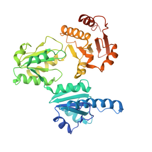
    Calsequestrin 1 is the principal Ca(2+) storage protein of the sarcoplasmic reticulum of skeletal muscle. Its inheritable D244G mutation causes a myopathy with vacuolar aggregates, whereas its M87T "variant" is weakly associated with malignant hyperthermia. We characterized the consequences of these mutations with studies of the human proteins in vitro. Equilibrium dialysis and turbidity measurements showed that D244G and, to a lesser extent, M87T partially lose Ca(2+) binding exhibited by wild type calsequestrin 1 at high Ca(2+) concentrations. D244G aggregates abruptly and abnormally, a property that fully explains the protein inclusions that characterize its phenotype. D244G crystallized in low Ca(2+) concentrations lacks two Ca(2+) ions normally present in wild type that weakens the hydrophobic core of Domain II. D244G crystallized in high Ca(2+) concentrations regains its missing ions and Domain II order but shows a novel dimeric interaction. The M87T mutation causes a major shift of the α-helix bearing the mutated residue, significantly weakening the back-to-back interface essential for tetramerization. D244G exhibited the more severe structural and biophysical property changes, which matches the different pathophysiological impacts of these mutations.


  • Organizational Affiliation
    • From the Department of Chemistry, Washington State University, Pullman, Washington 99164-4630.

Macromolecule Content 

  • Total Structure Weight: 41.98 kDa 
  • Atom Count: 2,700 
  • Modeled Residue Count: 347 
  • Deposited Residue Count: 362 
  • Unique protein chains: 1

Macromolecules

Find similar proteins by:|  3D Structure
Entity ID: 1
MoleculeChains  Sequence LengthOrganismDetailsImage
Calsequestrin-1362Homo sapiensMutation(s): 1 
Gene Names: CASQ1CASQ
UniProt & NIH Common Fund Data Resources
Find proteins for P31415 (Homo sapiens)
Explore P31415 
Go to UniProtKB:  P31415
PHAROS:  P31415
GTEx:  ENSG00000143318 
Entity Groups
Sequence Clusters30% Identity50% Identity70% Identity90% Identity95% Identity100% Identity
UniProt GroupP31415
Sequence Annotations
Expand
Reference Sequence

Experimental Data & Validation

Experimental Data

  • Method: X-RAY DIFFRACTION
  • Resolution: 3.31 Å
  • R-Value Free:  0.290 (Depositor), 0.289 (DCC) 
  • R-Value Work:  0.249 (Depositor), 0.251 (DCC) 
  • R-Value Observed: 0.253 (Depositor) 
Space Group: P 21 21 2
Unit Cell:
Length ( Å )Angle ( ˚ )
a = 66.106α = 90
b = 82.815β = 90
c = 89.269γ = 90
Software Package:
Software NamePurpose
PHENIXrefinement
HKL-2000data reduction
Cootmodel building
PHENIXphasing

Structure Validation

View Full Validation Report


Entry History 

Deposition Data

Revision History  (Full details and data files)

  • Version 1.0: 2015-10-07
    Type: Initial release
  • Version 1.1: 2015-10-28
    Changes: Data collection, Database references
  • Version 1.2: 2015-12-09
    Changes: Database references
  • Version 1.3: 2023-09-27
    Changes: Data collection, Database references, Derived calculations, Refinement description