5CPV | pdb_00005cpv

RESTRAINED LEAST SQUARES REFINEMENT OF NATIVE (CALCIUM) AND CADMIUM-SUBSTITUTED CARP PARVALBUMIN USING X-RAY CRYSTALLOGRAPHIC DATA AT 1.6-ANGSTROMS RESOLUTION


Experimental Data Snapshot

  • Method: X-RAY DIFFRACTION
  • Resolution: 1.60 Å
  • R-Value Observed: 
    0.187 (Depositor) 

wwPDB Validation 3D Report Full Report

Validation slider image for 5CPV

This is version 1.4 of the entry. See complete history

Literature

Restrained least squares refinement of native (calcium) and cadmium-substituted carp parvalbumin using X-ray crystallographic data at 1.6-A resolution.

Swain, A.L.Kretsinger, R.H.Amma, E.L.

(1989) J Biological Chem 264: 16620-16628

  • Primary Citation Related Structures: 
    1CDP, 5CPV

  • PubMed Abstract: 

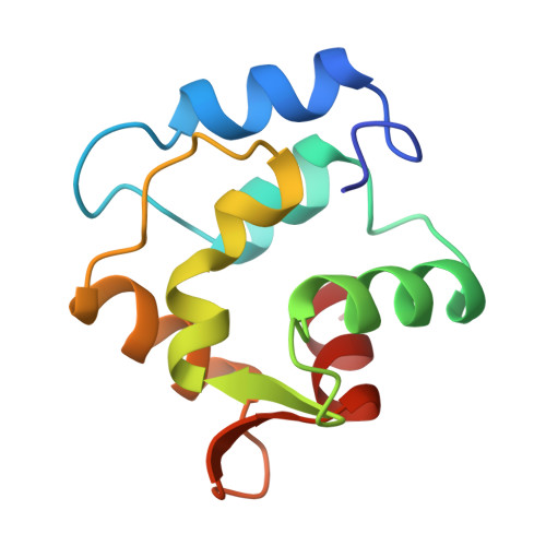
    Carp parvalbumin coordinates calcium through one carbonyl oxygen atom and the oxygen-containing side chains of 5 amino acid residues, or 4 residues and a water molecule, in a helix-loop-helix structural motif. Other calcium-binding proteins, including calmodulin and troponin C, also possess this unique calcium-binding design, which is designated EF-hand or calmodulin fold. Parvalbumin has two such sites, labeled CD and EF. Each of the calcium-binding sites of refined structures of proteins belonging to this group has a 7-oxygen coordination sphere except those of the structure of parvalbumin as it was reported in 1975. This structure had been refined at 1.9 A using difference Fourier techniques on film data. The CD site appeared to be 6-coordinate and the EF site 8-coordinate. Results of NMR experiments using 113Cd-substituted parvalbumin, however, indicate that the sites are similar to one another with coordination number greater than 6. To resolve the inconsistency between crystallographic and NMR results, 1.6 A area detector data was collected for native and cadmium-substituted parvalbumin; the structures have been refined to R factors of 18.7% and 16.4%, respectively, with acceptable geometry and low errors in atomic coordinates. Differences between the parvalbumin structure described in 1975 and the present structure are addressed, including the discovery of 7-coordination for both the CD and EF sites.


  • Organizational Affiliation
    • Department of Chemistry, University of South Carolina, Columbia 29208.

Macromolecule Content 

  • Total Structure Weight: 11.56 kDa 
  • Atom Count: 886 
  • Modeled Residue Count: 109 
  • Deposited Residue Count: 109 
  • Unique protein chains: 1

Macromolecules

Find similar proteins by:|  3D Structure
Entity ID: 1
MoleculeChains  Sequence LengthOrganismDetailsImage
CALCIUM-BINDING PARVALBUMIN B109Cyprinus carpioMutation(s): 0 
UniProt
Find proteins for P02618 (Cyprinus carpio)
Explore P02618 
Go to UniProtKB:  P02618
Entity Groups
Sequence Clusters30% Identity50% Identity70% Identity90% Identity95% Identity100% Identity
UniProt GroupP02618
Sequence Annotations
Expand
Reference Sequence

Small Molecules

Experimental Data & Validation

Experimental Data

  • Method: X-RAY DIFFRACTION
  • Resolution: 1.60 Å
  • R-Value Observed: 0.187 (Depositor) 
Space Group: C 1 2 1
Unit Cell:
Length ( Å )Angle ( ˚ )
a = 28.7α = 90
b = 60.61β = 94.6
c = 54.35γ = 90
Software Package:
Software NamePurpose
PROLSQrefinement

Structure Validation

View Full Validation Report


Entry History 

Deposition Data

Revision History  (Full details and data files)

  • Version 1.0: 1990-10-15
    Type: Initial release
  • Version 1.1: 2008-03-03
    Changes: Version format compliance
  • Version 1.2: 2011-07-13
    Changes: Version format compliance
  • Version 1.3: 2017-11-29
    Changes: Derived calculations, Other
  • Version 1.4: 2024-11-06
    Changes: Data collection, Database references, Derived calculations, Structure summary