5CMC | pdb_00005cmc

Mnemiopsis leidyi ML032222a iGluR LBD E423S mutant glycine complex


Experimental Data Snapshot

  • Method: X-RAY DIFFRACTION
  • Resolution: 1.28 Å
  • R-Value Free: 
    0.157 (Depositor), 0.159 (DCC) 
  • R-Value Work: 
    0.134 (Depositor), 0.136 (DCC) 
  • R-Value Observed: 
    0.136 (Depositor) 

Starting Model: experimental
View more details

wwPDB Validation 3D Report Full Report

Validation slider image for 5CMC

This is version 1.4 of the entry. See complete history

Literature

Molecular lock regulates binding of glycine to a primitive NMDA receptor.

Yu, A.Alberstein, R.Thomas, A.Zimmet, A.Grey, R.Mayer, M.L.Lau, A.Y.

(2016) Proc Natl Acad Sci U S A 113: E6786-E6795

  • DOI: https://doi.org/10.1073/pnas.1607010113
  • Primary Citation Related Structures: 
    5CMB, 5CMC

  • PubMed Abstract: 

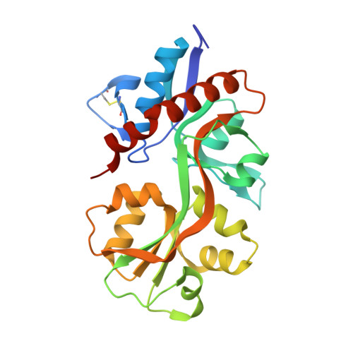
    The earliest metazoan ancestors of humans include the ctenophore Mnemiopsis leidyi The genome of this comb jelly encodes homologs of vertebrate ionotropic glutamate receptors (iGluRs) that are distantly related to glycine-activated NMDA receptors and that bind glycine with unusually high affinity. Using ligand-binding domain (LBD) mutants for electrophysiological analysis, we demonstrate that perturbing a ctenophore-specific interdomain Arg-Glu salt bridge that is notably absent from vertebrate AMPA, kainate, and NMDA iGluRs greatly increases the rate of recovery from desensitization, while biochemical analysis reveals a large decrease in affinity for glycine. X-ray crystallographic analysis details rearrangements in the binding pocket stemming from the mutations, and molecular dynamics simulations suggest that the interdomain salt bridge acts as a steric barrier regulating ligand binding and that the free energy required to access open conformations in the glycine-bound LBD is largely responsible for differences in ligand affinity among the LBD variants.


  • Organizational Affiliation
    • Department of Biophysics and Biophysical Chemistry, The Johns Hopkins University School of Medicine, Baltimore, MD 21205.

Macromolecule Content 

  • Total Structure Weight: 58.11 kDa 
  • Atom Count: 4,897 
  • Modeled Residue Count: 502 
  • Deposited Residue Count: 512 
  • Unique protein chains: 1

Macromolecules

Find similar proteins by:|  3D Structure
Entity ID: 1
MoleculeChains  Sequence LengthOrganismDetailsImage
ML032222a iGluR
A, B
256Mnemiopsis leidyiMutation(s): 0 
Gene Names: ML032222a
UniProt
Find proteins for A0A0R4I973 (Mnemiopsis leidyi)
Explore A0A0R4I973 
Go to UniProtKB:  A0A0R4I973
Entity Groups
Sequence Clusters30% Identity50% Identity70% Identity90% Identity95% Identity100% Identity
UniProt GroupA0A0R4I973
Sequence Annotations
Expand
Reference Sequence

Experimental Data & Validation

Experimental Data

  • Method: X-RAY DIFFRACTION
  • Resolution: 1.28 Å
  • R-Value Free:  0.157 (Depositor), 0.159 (DCC) 
  • R-Value Work:  0.134 (Depositor), 0.136 (DCC) 
  • R-Value Observed: 0.136 (Depositor) 
Space Group: P 1 21 1
Unit Cell:
Length ( Å )Angle ( ˚ )
a = 45.899α = 90
b = 123.138β = 111.72
c = 54.507γ = 90
Software Package:
Software NamePurpose
PHENIXrefinement
HKL-2000data reduction
HKL-2000data scaling
PHENIXphasing

Structure Validation

View Full Validation Report


Entry History 

Deposition Data

Revision History  (Full details and data files)

  • Version 1.0: 2016-07-20
    Type: Initial release
  • Version 1.1: 2016-11-09
    Changes: Database references
  • Version 1.2: 2016-11-16
    Changes: Database references
  • Version 1.3: 2023-09-27
    Changes: Data collection, Database references, Derived calculations, Refinement description
  • Version 1.4: 2024-10-23
    Changes: Structure summary