5CJ3 | pdb_00005cj3

Crystal structure of the zorbamycin binding protein (ZbmA) from Streptomyces flavoviridis with zorbamycin


Experimental Data Snapshot

  • Method: X-RAY DIFFRACTION
  • Resolution: 1.65 Å
  • R-Value Free: 
    0.213 (Depositor), 0.212 (DCC) 
  • R-Value Work: 
    0.176 (Depositor), 0.176 (DCC) 
  • R-Value Observed: 
    0.178 (Depositor) 

Starting Model: experimental
View more details

wwPDB Validation 3D Report Full Report

Validation slider image for 5CJ3

Ligand Structure Quality Assessment 


This is version 1.4 of the entry. See complete history

Literature

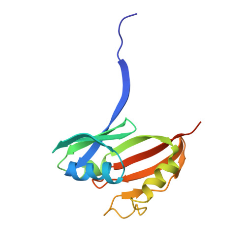
Crystal Structure of the Zorbamycin-Binding Protein ZbmA, the Primary Self-Resistance Element in Streptomyces flavoviridis ATCC21892.

Rudolf, J.D.Bigelow, L.Chang, C.Cuff, M.E.Lohman, J.R.Chang, C.Y.Ma, M.Yang, D.Clancy, S.Babnigg, G.Joachimiak, A.Phillips, G.N.Shen, B.

(2015) Biochemistry 54: 6842-6851

  • DOI: https://doi.org/10.1021/acs.biochem.5b01008
  • Primary Citation Related Structures: 
    4IAG, 5CJ3

  • PubMed Abstract: 

    The bleomycins (BLMs), tallysomycins (TLMs), phleomycin, and zorbamycin (ZBM) are members of the BLM family of glycopeptide-derived antitumor antibiotics. The BLM-producing Streptomyces verticillus ATCC15003 and the TLM-producing Streptoalloteichus hindustanus E465-94 ATCC31158 both possess at least two self-resistance elements, an N-acetyltransferase and a binding protein. The N-acetyltransferase provides resistance by disrupting the metal-binding domain of the antibiotic that is required for activity, while the binding protein confers resistance by sequestering the metal-bound antibiotic and preventing drug activation via molecular oxygen. We recently established that the ZBM producer, Streptomyces flavoviridis ATCC21892, lacks the N-acetyltransferase resistance gene and that the ZBM-binding protein, ZbmA, is sufficient to confer resistance in the producing strain. To investigate the resistance mechanism attributed to ZbmA, we determined the crystal structures of apo and Cu(II)-ZBM-bound ZbmA at high resolutions of 1.90 and 1.65 Å, respectively. A comparison and contrast with other structurally characterized members of the BLM-binding protein family revealed key differences in the protein-ligand binding environment that fine-tunes the ability of ZbmA to sequester metal-bound ZBM and supports drug sequestration as the primary resistance mechanism in the producing organisms of the BLM family of antitumor antibiotics.


  • Organizational Affiliation
    • Department of Chemistry, The Scripps Research Institute , Jupiter, Florida 33458, United States.

Macromolecule Content 

  • Total Structure Weight: 32.78 kDa 
  • Atom Count: 2,575 
  • Modeled Residue Count: 252 
  • Deposited Residue Count: 270 
  • Unique protein chains: 1

Macromolecules

Find similar proteins by:|  3D Structure
Entity ID: 1
MoleculeChains  Sequence LengthOrganismDetailsImage
Zbm binding protein
A, B
135Streptomyces pilosusMutation(s): 0 
Gene Names: zbmA
UniProt
Find proteins for B9UIZ4 (Streptomyces pilosus)
Explore B9UIZ4 
Go to UniProtKB:  B9UIZ4
Entity Groups
Sequence Clusters30% Identity50% Identity70% Identity90% Identity95% Identity100% Identity
UniProt GroupB9UIZ4
Sequence Annotations
Expand
Reference Sequence

Experimental Data & Validation

Experimental Data

  • Method: X-RAY DIFFRACTION
  • Resolution: 1.65 Å
  • R-Value Free:  0.213 (Depositor), 0.212 (DCC) 
  • R-Value Work:  0.176 (Depositor), 0.176 (DCC) 
  • R-Value Observed: 0.178 (Depositor) 
Space Group: P 21 21 21
Unit Cell:
Length ( Å )Angle ( ˚ )
a = 40.737α = 90
b = 78.989β = 90
c = 79.542γ = 90
Software Package:
Software NamePurpose
SCALEPACKdata scaling
PHENIXrefinement
PDB_EXTRACTdata extraction
HKL-3000data reduction
HKL-3000phasing
SBC-Collectdata collection

Structure Validation

View Full Validation Report


Ligand Structure Quality Assessment 


Entry History 

Revision History  (Full details and data files)

  • Version 1.0: 2015-07-22
    Type: Initial release
  • Version 1.1: 2015-11-11
    Changes: Database references
  • Version 1.2: 2015-11-25
    Changes: Database references
  • Version 1.3: 2023-09-27
    Changes: Data collection, Database references, Derived calculations, Refinement description
  • Version 1.4: 2025-10-22
    Changes: Structure summary