5CH7 | pdb_00005ch7

Crystal structure of the perchlorate reductase PcrAB - Phe164 gate switch intermediate - from Azospira suillum PS


Experimental Data Snapshot

  • Method: X-RAY DIFFRACTION
  • Resolution: 2.20 Å
  • R-Value Free: 
    0.233 (Depositor), 0.191 (DCC) 
  • R-Value Work: 
    0.178 (Depositor) 
  • R-Value Observed: 
    0.181 (Depositor) 

Starting Model: experimental
View more details

wwPDB Validation 3D Report Full Report

Validation slider image for 5CH7

Ligand Structure Quality Assessment 


This is version 1.5 of the entry. See complete history

Literature

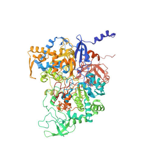
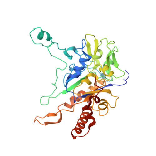
Perchlorate Reductase Is Distinguished by Active Site Aromatic Gate Residues.

Youngblut, M.D.Tsai, C.L.Clark, I.C.Carlson, H.K.Maglaqui, A.P.Gau-Pan, P.S.Redford, S.A.Wong, A.Tainer, J.A.Coates, J.D.

(2016) J Biological Chem 291: 9190-9202

  • DOI: https://doi.org/10.1074/jbc.M116.714618
  • Primary Citation Related Structures: 
    4YDD, 5CH7, 5CHC, 5E7O

  • PubMed Abstract: 

    Perchlorate is an important ion on both Earth and Mars. Perchlorate reductase (PcrAB), a specialized member of the dimethylsulfoxide reductase superfamily, catalyzes the first step of microbial perchlorate respiration, but little is known about the biochemistry, specificity, structure, and mechanism of PcrAB. Here we characterize the biophysics and phylogeny of this enzyme and report the 1.86-Å resolution PcrAB complex crystal structure. Biochemical analysis revealed a relatively high perchlorate affinity (Km = 6 μm) and a characteristic substrate inhibition compared with the highly similar respiratory nitrate reductase NarGHI, which has a relatively much lower affinity for perchlorate (Km = 1.1 mm) and no substrate inhibition. Structural analysis of oxidized and reduced PcrAB with and without the substrate analog SeO3 (2-) bound to the active site identified key residues in the positively charged and funnel-shaped substrate access tunnel that gated substrate entrance and product release while trapping transiently produced chlorate. The structures suggest gating was associated with shifts of a Phe residue between open and closed conformations plus an Asp residue carboxylate shift between monodentate and bidentate coordination to the active site molybdenum atom. Taken together, structural and mutational analyses of gate residues suggest key roles of these gate residues for substrate entrance and product release. Our combined results provide the first detailed structural insight into the mechanism of biological perchlorate reduction, a critical component of the chlorine redox cycle on Earth.


  • Organizational Affiliation
    • From the Energy Biosciences Institute and.

Macromolecule Content 

  • Total Structure Weight: 429.08 kDa 
  • Atom Count: 31,660 
  • Modeled Residue Count: 3,661 
  • Deposited Residue Count: 3,696 
  • Unique protein chains: 2

Macromolecules

Find similar proteins by:|  3D Structure
Entity ID: 1
MoleculeChains  Sequence LengthOrganismDetailsImage
DMSO reductase family type II enzyme, molybdopterin subunit
A, C, E
899Azospira oryzae PSMutation(s): 0 
UniProt
Find proteins for G8QM55 (Azospira oryzae (strain ATCC BAA-33 / DSM 13638 / PS))
Explore G8QM55 
Go to UniProtKB:  G8QM55
Entity Groups
Sequence Clusters30% Identity50% Identity70% Identity90% Identity95% Identity100% Identity
UniProt GroupG8QM55
Sequence Annotations
Expand
Reference Sequence
Find similar proteins by:|  3D Structure
Entity ID: 2
MoleculeChains  Sequence LengthOrganismDetailsImage
DMSO reductase family type II enzyme, iron-sulfur subunit
B, D, F
333Azospira oryzae PSMutation(s): 0 
UniProt
Find proteins for G8QM54 (Azospira oryzae (strain ATCC BAA-33 / DSM 13638 / PS))
Explore G8QM54 
Go to UniProtKB:  G8QM54
Entity Groups
Sequence Clusters30% Identity50% Identity70% Identity90% Identity95% Identity100% Identity
UniProt GroupG8QM54
Sequence Annotations
Expand
Reference Sequence

Small Molecules

Ligands 11 Unique
IDChains Name / Formula / InChI Key2D Diagram3D Interactions
MD1

Query on MD1


Download:Ideal Coordinates CCD File
DA [auth C],
J [auth A],
UA [auth E]
PHOSPHORIC ACID 4-(2-AMINO-4-OXO-3,4,5,6,-TETRAHYDRO-PTERIDIN-6-YL)-2-HYDROXY-3,4-DIMERCAPTO-BUT-3-EN-YL ESTER GUANYLATE ESTER
C20 H26 N10 O13 P2 S2
IRGDLSAXQOKWLX-XHEYTWMPSA-N
MGD

Query on MGD


Download:Ideal Coordinates CCD File
CA [auth C],
I [auth A],
TA [auth E]
2-AMINO-5,6-DIMERCAPTO-7-METHYL-3,7,8A,9-TETRAHYDRO-8-OXA-1,3,9,10-TETRAAZA-ANTHRACEN-4-ONE GUANOSINE DINUCLEOTIDE
C20 H26 N10 O13 P2 S2
VQAGYJCYOLHZDH-ILXWUORBSA-N
SF4

Query on SF4


Download:Ideal Coordinates CCD File
AA [auth C]
CB [auth F]
DB [auth F]
EB [auth F]
G [auth A]
AA [auth C],
CB [auth F],
DB [auth F],
EB [auth F],
G [auth A],
NA [auth D],
OA [auth D],
PA [auth D],
RA [auth E],
U [auth B],
V [auth B],
W [auth B]
IRON/SULFUR CLUSTER
Fe4 S4
LJBDFODJNLIPKO-UHFFFAOYSA-N
F3S

Query on F3S


Download:Ideal Coordinates CCD File
BB [auth F],
MA [auth D],
T [auth B]
FE3-S4 CLUSTER
Fe3 S4
FCXHZBQOKRZXKS-UHFFFAOYSA-N
MO

Query on MO


Download:Ideal Coordinates CCD File
BA [auth C],
H [auth A],
SA [auth E]
MOLYBDENUM ATOM
Mo
ZOKXTWBITQBERF-UHFFFAOYSA-N
GOL

Query on GOL


Download:Ideal Coordinates CCD File
P [auth A]GLYCEROL
C3 H8 O3
PEDCQBHIVMGVHV-UHFFFAOYSA-N
SO3

Query on SO3


Download:Ideal Coordinates CCD File
IA [auth C],
JA [auth C],
Q [auth A],
S [auth A],
YA [auth E]
SULFITE ION
O3 S
LSNNMFCWUKXFEE-UHFFFAOYSA-L
ZN

Query on ZN


Download:Ideal Coordinates CCD File
AB [auth E],
KA [auth C],
O [auth A]
ZINC ION
Zn
PTFCDOFLOPIGGS-UHFFFAOYSA-N
EDO

Query on EDO


Download:Ideal Coordinates CCD File
EA [auth C]
FA [auth C]
FB [auth F]
GB [auth F]
K [auth A]
EA [auth C],
FA [auth C],
FB [auth F],
GB [auth F],
K [auth A],
L [auth A],
LA [auth D],
M [auth A],
QA [auth D],
R [auth A],
VA [auth E],
WA [auth E],
X [auth B],
XA [auth E],
Y [auth B],
Z [auth B]
1,2-ETHANEDIOL
C2 H6 O2
LYCAIKOWRPUZTN-UHFFFAOYSA-N
ACT

Query on ACT


Download:Ideal Coordinates CCD File
HA [auth C]ACETATE ION
C2 H3 O2
QTBSBXVTEAMEQO-UHFFFAOYSA-M
NA

Query on NA


Download:Ideal Coordinates CCD File
GA [auth C],
HB [auth F],
N [auth A],
ZA [auth E]
SODIUM ION
Na
FKNQFGJONOIPTF-UHFFFAOYSA-N

Experimental Data & Validation

Experimental Data

  • Method: X-RAY DIFFRACTION
  • Resolution: 2.20 Å
  • R-Value Free:  0.233 (Depositor), 0.191 (DCC) 
  • R-Value Work:  0.178 (Depositor) 
  • R-Value Observed: 0.181 (Depositor) 
Space Group: P 21 21 21
Unit Cell:
Length ( Å )Angle ( ˚ )
a = 132.884α = 90
b = 175.667β = 90
c = 193.278γ = 90
Software Package:
Software NamePurpose
PHENIXrefinement
Aimlessdata scaling
PHASERphasing

Structure Validation

View Full Validation Report


Ligand Structure Quality Assessment 


Entry History 

Deposition Data

Revision History  (Full details and data files)

  • Version 1.0: 2016-03-09
    Type: Initial release
  • Version 1.1: 2016-03-16
    Changes: Database references
  • Version 1.2: 2016-05-11
    Changes: Database references
  • Version 1.3: 2017-11-01
    Changes: Author supporting evidence, Database references, Derived calculations
  • Version 1.4: 2024-03-06
    Changes: Data collection, Database references, Derived calculations
  • Version 1.5: 2026-03-04
    Changes: Refinement description, Structure summary