5C7Q | pdb_00005c7q

Crystal Structure of the Bdellovibrio bacteriovorus Nucleoside Diphosphate Sugar Hydrolase


Experimental Data Snapshot

  • Method: X-RAY DIFFRACTION
  • Resolution: 1.52 Å
  • R-Value Free: 
    0.215 (Depositor), 0.223 (DCC) 
  • R-Value Work: 
    0.186 (Depositor), 0.198 (DCC) 
  • R-Value Observed: 
    0.188 (Depositor) 

wwPDB Validation 3D Report Full Report

Validation slider image for 5C7Q

This is version 1.1 of the entry. See complete history

Literature

Structural and Enzymatic Characterization of a Nucleoside Diphosphate Sugar Hydrolase from Bdellovibrio bacteriovorus.

de la Pena, A.H.Suarez, A.Duong-Ly, K.C.Schoeffield, A.J.Pizarro-Dupuy, M.A.Zarr, M.Pineiro, S.A.Amzel, L.M.Gabelli, S.B.

(2015) PLoS One 10: e0141716-e0141716

  • DOI: https://doi.org/10.1371/journal.pone.0141716
  • Primary Citation Related Structures: 
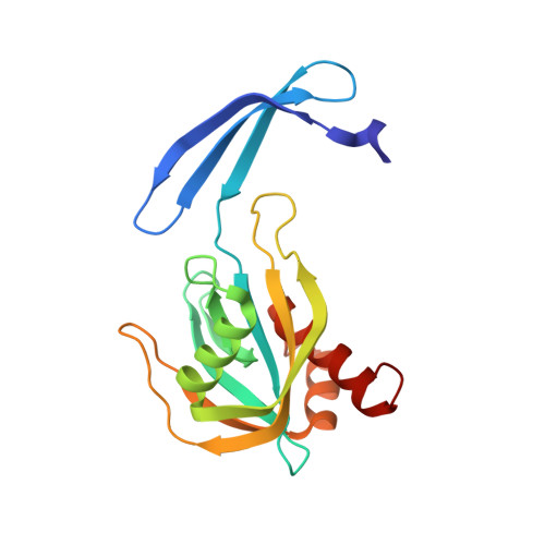
    5C7Q, 5C7T, 5C8L

  • PubMed Abstract: 

    Given the broad range of substrates hydrolyzed by Nudix (nucleoside diphosphate linked to X) enzymes, identification of sequence and structural elements that correctly predict a Nudix substrate or characterize a family is key to correctly annotate the myriad of Nudix enzymes. Here, we present the structure determination and characterization of Bd3179 -- a Nudix hydrolase from Bdellovibrio bacteriovorus-that we show localized in the periplasmic space of this obligate Gram-negative predator. We demonstrate that the enzyme is a nucleoside diphosphate sugar hydrolase (NDPSase) and has a high degree of sequence and structural similarity to a canonical ADP-ribose hydrolase and to a nucleoside diphosphate sugar hydrolase (1.4 and 1.3 Å Cα RMSD respectively). Examination of the structural elements conserved in both types of enzymes confirms that an aspartate-X-lysine motif on the C-terminal helix of the α-β-α NDPSase fold differentiates NDPSases from ADPRases.


  • Organizational Affiliation
    • Department of Biomedical Engineering, Johns Hopkins University School of Medicine, Baltimore, Maryland, United States of America; Structural Enzymology and Thermodynamics Group, Department of Biophysics and Biophysical Chemistry, Johns Hopkins University School of Medicine, Baltimore, Maryland, United States of America.

Macromolecule Content 

  • Total Structure Weight: 44.74 kDa 
  • Atom Count: 3,497 
  • Modeled Residue Count: 362 
  • Deposited Residue Count: 364 
  • Unique protein chains: 1

Macromolecules

Find similar proteins by:|  3D Structure
Entity ID: 1
MoleculeChains  Sequence LengthOrganismDetailsImage
NudF protein
A, B
182Bdellovibrio bacteriovorus HD100Mutation(s): 0 
Gene Names: nudFBd3179
EC: 3.6.1.13
UniProt
Find proteins for Q6MIH8 (Bdellovibrio bacteriovorus (strain ATCC 15356 / DSM 50701 / NCIMB 9529 / HD100))
Explore Q6MIH8 
Go to UniProtKB:  Q6MIH8
Entity Groups
Sequence Clusters30% Identity50% Identity70% Identity90% Identity95% Identity100% Identity
UniProt GroupQ6MIH8
Sequence Annotations
Expand
Reference Sequence

Small Molecules

Ligands 5 Unique
IDChains Name / Formula / InChI Key2D Diagram3D Interactions
PGE

Query on PGE


Download:Ideal Coordinates CCD File
M [auth A],
W [auth B]
TRIETHYLENE GLYCOL
C6 H14 O4
ZIBGPFATKBEMQZ-UHFFFAOYSA-N
PG0

Query on PG0


Download:Ideal Coordinates CCD File
L [auth A],
V [auth B]
2-(2-METHOXYETHOXY)ETHANOL
C5 H12 O3
SBASXUCJHJRPEV-UHFFFAOYSA-N
PEG

Query on PEG


Download:Ideal Coordinates CCD File
J [auth A],
K [auth A]
DI(HYDROXYETHYL)ETHER
C4 H10 O3
MTHSVFCYNBDYFN-UHFFFAOYSA-N
SO4

Query on SO4


Download:Ideal Coordinates CCD File
C [auth A]
D [auth A]
E [auth A]
N [auth A]
O [auth A]
C [auth A],
D [auth A],
E [auth A],
N [auth A],
O [auth A],
P [auth B]
SULFATE ION
O4 S
QAOWNCQODCNURD-UHFFFAOYSA-L
GOL

Query on GOL


Download:Ideal Coordinates CCD File
F [auth A]
G [auth A]
H [auth A]
I [auth A]
Q [auth B]
F [auth A],
G [auth A],
H [auth A],
I [auth A],
Q [auth B],
R [auth B],
S [auth B],
T [auth B],
U [auth B]
GLYCEROL
C3 H8 O3
PEDCQBHIVMGVHV-UHFFFAOYSA-N

Experimental Data & Validation

Experimental Data

  • Method: X-RAY DIFFRACTION
  • Resolution: 1.52 Å
  • R-Value Free:  0.215 (Depositor), 0.223 (DCC) 
  • R-Value Work:  0.186 (Depositor), 0.198 (DCC) 
  • R-Value Observed: 0.188 (Depositor) 
Space Group: P 21 21 2
Unit Cell:
Length ( Å )Angle ( ˚ )
a = 75.425α = 90
b = 103.098β = 90
c = 51.712γ = 90
Software Package:
Software NamePurpose
DENZOdata reduction
SCALEPACKdata scaling
REFMACrefinement
PDB_EXTRACTdata extraction
HKL-2000data reduction
SOLVEphasing

Structure Validation

View Full Validation Report


Entry History 

Deposition Data

Revision History  (Full details and data files)

  • Version 1.0: 2016-01-20
    Type: Initial release
  • Version 1.1: 2024-03-06
    Changes: Data collection, Database references, Derived calculations