5C6R | pdb_00005c6r

Crystal structure of PH domain of ASAP1


Experimental Data Snapshot

  • Method: X-RAY DIFFRACTION
  • Resolution: 1.80 Å
  • R-Value Free: 
    0.260 (Depositor), 0.256 (DCC) 
  • R-Value Work: 
    0.208 (Depositor), 0.214 (DCC) 
  • R-Value Observed: 
    0.211 (Depositor) 

Starting Model: experimental
View more details

wwPDB Validation 3D Report Full Report

Validation slider image for 5C6R

This is version 1.2 of the entry. See complete history

Literature

Molecular Basis for Cooperative Binding of Anionic Phospholipids to the PH Domain of the Arf GAP ASAP1.

Jian, X.Tang, W.K.Zhai, P.Roy, N.S.Luo, R.Gruschus, J.M.Yohe, M.E.Chen, P.W.Li, Y.Byrd, R.A.Xia, D.Randazzo, P.A.

(2015) Structure 23: 1977-1988

  • DOI: https://doi.org/10.1016/j.str.2015.08.008
  • Primary Citation Related Structures: 
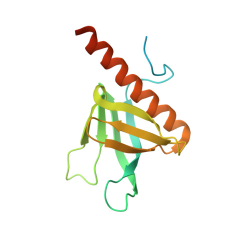
    5C6R, 5C79

  • PubMed Abstract: 

    We have defined the molecular basis for association of the PH domain of the Arf GAP ASAP1 with phospholipid bilayers. Structures of the unliganded and dibutyryl PtdIns(4,5)P2-bound PH domain were solved. PtdIns(4,5)P2 made contact with both a canonical site (C site) and an atypical site (A site). We hypothesized cooperative binding of PtdIns(4,5)P2 to the C site and a nonspecific anionic phospholipid to the A site. PtdIns(4,5)P2 dependence of binding to large unilamellar vesicles and GAP activity was sigmoidal, consistent with cooperative sites. In contrast, PtdIns(4,5)P2 binding to the PH domain of PLC δ1 was hyperbolic. Mutation of amino acids in either the C or A site resulted in decreased PtdIns(4,5)P2-dependent binding to vesicles and decreased GAP activity. The results support the idea of cooperative phospholipid binding to the C and A sites of the PH domain of ASAP1. We propose that the mechanism underlies rapid switching between active and inactive ASAP1.


  • Organizational Affiliation
    • Laboratory of Cellular and Molecular Biology, National Cancer Institute, National Institutes of Health, Bethesda, MD 20892, USA.

Macromolecule Content 

  • Total Structure Weight: 35.15 kDa 
  • Atom Count: 1,838 
  • Modeled Residue Count: 218 
  • Deposited Residue Count: 300 
  • Unique protein chains: 1

Macromolecules

Find similar proteins by:|  3D Structure
Entity ID: 1
MoleculeChains  Sequence LengthOrganismDetailsImage
Arf-GAP
A, B
150Mus musculusMutation(s): 0 
Gene Names: Asap1Ddef1Kiaa1249Shag1
UniProt & NIH Common Fund Data Resources
Find proteins for Q9QWY8 (Mus musculus)
Explore Q9QWY8 
Go to UniProtKB:  Q9QWY8
IMPC:  MGI:1342335
Entity Groups
Sequence Clusters30% Identity50% Identity70% Identity90% Identity95% Identity100% Identity
UniProt GroupQ9QWY8
Sequence Annotations
Expand
Reference Sequence

Experimental Data & Validation

Experimental Data

  • Method: X-RAY DIFFRACTION
  • Resolution: 1.80 Å
  • R-Value Free:  0.260 (Depositor), 0.256 (DCC) 
  • R-Value Work:  0.208 (Depositor), 0.214 (DCC) 
  • R-Value Observed: 0.211 (Depositor) 
Space Group: P 43
Unit Cell:
Length ( Å )Angle ( ˚ )
a = 48.12α = 90
b = 48.12β = 90
c = 111.333γ = 90
Software Package:
Software NamePurpose
REFMACrefinement
DENZOdata reduction
SCALEPACKdata scaling
MOLREPphasing

Structure Validation

View Full Validation Report


Entry History 

Deposition Data

Revision History  (Full details and data files)

  • Version 1.0: 2015-10-07
    Type: Initial release
  • Version 1.1: 2015-11-18
    Changes: Database references
  • Version 1.2: 2023-09-27
    Changes: Data collection, Database references, Derived calculations, Refinement description