5C2I | pdb_00005c2i

Crystal structure of Anabaena sp. DyP-type peroxidese (AnaPX)


Experimental Data Snapshot

  • Method: X-RAY DIFFRACTION
  • Resolution: 1.89 Å
  • R-Value Free: 
    0.199 (Depositor), 0.208 (DCC) 
  • R-Value Work: 
    0.181 (Depositor), 0.190 (DCC) 
  • R-Value Observed: 
    0.182 (Depositor) 

Starting Model: experimental
View more details

wwPDB Validation 3D Report Full Report

Validation slider image for 5C2I

Ligand Structure Quality Assessment 


This is version 1.4 of the entry. See complete history

Literature

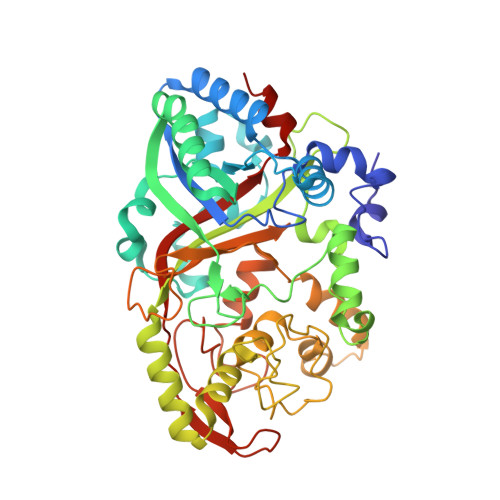
Anabaena sp. DyP-type peroxidase is a tetramer consisting of two asymmetric dimers.

Yoshida, T.Ogola, H.J.Amano, Y.Hisabori, T.Ashida, H.Sawa, Y.Tsuge, H.Sugano, Y.

(2016) Proteins 84: 31-42

  • DOI: https://doi.org/10.1002/prot.24952
  • Primary Citation Related Structures: 
    5C2I

  • PubMed Abstract: 

    DyP-type peroxidases are a newly discovered family of heme peroxidases distributed from prokaryotes to eukaryotes. Recently, using a structure-based sequence alignment, we proposed the new classes, P, I and V, as substitutes for classes A, B, C, and D [Arch Biochem Biophys 2015;574:49-55]. Although many class V enzymes from eukaryotes have been characterized, only two from prokaryotes have been reported. Here, we show the crystal structure of one of these two enzymes, Anabaena sp. DyP-type peroxidase (AnaPX). AnaPX is tetramer formed from Cys224-Cys224 disulfide-linked dimers. The tetramer of wild-type AnaPX was stable at all salt concentrations tested. In contrast, the C224A mutant showed salt concentration-dependent oligomeric states: in 600 mM NaCl, it maintained a tetrameric structure, whereas in the absence of salt, it dissociated into monomers, leading to a reduction in thermostability. Although the tetramer exhibits non-crystallographic, 2-fold symmetry in the asymmetric unit, two subunits forming the Cys224-Cys224 disulfide-linked dimer are related by 165° rotation. This asymmetry creates an opening to cavities facing the inside of the tetramer, providing a pathway for hydrogen peroxide access. Finally, a phylogenetic analysis using structure-based sequence alignments showed that class V enzymes from prokaryotes, including AnaPX, are phylogenetically closely related to class V enzymes from eukaryotes.


  • Organizational Affiliation
    • Department of Bioresource and Environmental Sciences, Faculty of Life Sciences, Kyoto Sangyo University, Kamigamo-Motoyama, Kyoto, 603-8555, Japan.

Macromolecule Content 

  • Total Structure Weight: 216.94 kDa 
  • Atom Count: 16,111 
  • Modeled Residue Count: 1,850 
  • Deposited Residue Count: 1,872 
  • Unique protein chains: 1

Macromolecules

Find similar proteins by:|  3D Structure
Entity ID: 1
MoleculeChains  Sequence LengthOrganismDetailsImage
Alr1585 protein
A, B, C, D
468Nostoc sp. PCC 7120 = FACHB-418Mutation(s): 1 
Gene Names: alr1585
UniProt
Find proteins for A0ACD6B9A9 (Nostoc sp. (strain PCC 7120 / SAG 25.82 / UTEX 2576))
Explore A0ACD6B9A9 
Go to UniProtKB:  A0ACD6B9A9
Entity Groups
Sequence Clusters30% Identity50% Identity70% Identity90% Identity95% Identity100% Identity
UniProt GroupA0ACD6B9A9
Sequence Annotations
Expand
Reference Sequence

Small Molecules

Ligands 4 Unique
IDChains Name / Formula / InChI Key2D Diagram3D Interactions
HEM

Query on HEM


Download:Ideal Coordinates CCD File
E [auth A],
H [auth B],
J [auth C],
N [auth D]
PROTOPORPHYRIN IX CONTAINING FE
C34 H32 Fe N4 O4
KABFMIBPWCXCRK-RGGAHWMASA-L
GOL

Query on GOL


Download:Ideal Coordinates CCD File
F [auth A],
I [auth B],
K [auth C],
O [auth D],
P [auth D]
GLYCEROL
C3 H8 O3
PEDCQBHIVMGVHV-UHFFFAOYSA-N
EDO

Query on EDO


Download:Ideal Coordinates CCD File
G [auth A],
M [auth C]
1,2-ETHANEDIOL
C2 H6 O2
LYCAIKOWRPUZTN-UHFFFAOYSA-N
ACY

Query on ACY


Download:Ideal Coordinates CCD File
L [auth C]ACETIC ACID
C2 H4 O2
QTBSBXVTEAMEQO-UHFFFAOYSA-N

Experimental Data & Validation

Experimental Data

  • Method: X-RAY DIFFRACTION
  • Resolution: 1.89 Å
  • R-Value Free:  0.199 (Depositor), 0.208 (DCC) 
  • R-Value Work:  0.181 (Depositor), 0.190 (DCC) 
  • R-Value Observed: 0.182 (Depositor) 
Space Group: P 21 21 21
Unit Cell:
Length ( Å )Angle ( ˚ )
a = 105.734α = 90
b = 132.601β = 90
c = 176.528γ = 90
Software Package:
Software NamePurpose
HKL-2000data collection
HKL-2000data scaling
MOLREPphasing
REFMACrefinement
PDB_EXTRACTdata extraction

Structure Validation

View Full Validation Report


Ligand Structure Quality Assessment 


Entry History 

& Funding Information

Deposition Data


Funding OrganizationLocationGrant Number
JSPSJapan26450132

Revision History  (Full details and data files)

  • Version 1.0: 2015-12-16
    Type: Initial release
  • Version 1.1: 2016-01-27
    Changes: Database references
  • Version 1.2: 2020-02-19
    Changes: Data collection, Database references, Derived calculations
  • Version 1.3: 2023-11-08
    Changes: Data collection, Database references, Derived calculations, Refinement description
  • Version 1.4: 2024-11-13
    Changes: Structure summary