5BZV | pdb_00005bzv

Crystal structure of the RNA-binding domain of yeast Puf5p bound to SMX2 RNA


Experimental Data Snapshot

  • Method: X-RAY DIFFRACTION
  • Resolution: 2.35 Å
  • R-Value Free: 
    0.233 (Depositor), 0.234 (DCC) 
  • R-Value Work: 
    0.188 (Depositor), 0.191 (DCC) 
  • R-Value Observed: 
    0.192 (Depositor) 

Starting Model: experimental
View more details

wwPDB Validation 3D Report Full Report

Validation slider image for 5BZV

This is version 1.2 of the entry. See complete history

Literature

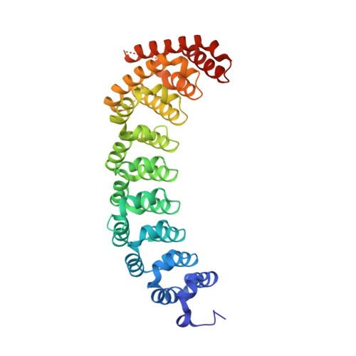
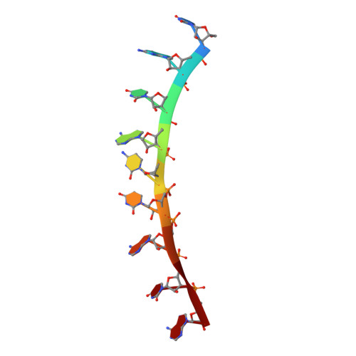
RNA regulatory networks diversified through curvature of the PUF protein scaffold.

Wilinski, D.Qiu, C.Lapointe, C.P.Nevil, M.Campbell, Z.T.Tanaka Hall, T.M.Wickens, M.

(2015) Nat Commun 6: 8213-8213

  • DOI: https://doi.org/10.1038/ncomms9213
  • Primary Citation Related Structures: 
    5BYM, 5BZ1, 5BZ5, 5BZU, 5BZV

  • PubMed Abstract: 

    Proteins bind and control mRNAs, directing their localization, translation and stability. Members of the PUF family of RNA-binding proteins control multiple mRNAs in a single cell, and play key roles in development, stem cell maintenance and memory formation. Here we identified the mRNA targets of a S. cerevisiae PUF protein, Puf5p, by ultraviolet-crosslinking-affinity purification and high-throughput sequencing (HITS-CLIP). The binding sites recognized by Puf5p are diverse, with variable spacer lengths between two specific sequences. Each length of site correlates with a distinct biological function. Crystal structures of Puf5p-RNA complexes reveal that the protein scaffold presents an exceptionally flat and extended interaction surface relative to other PUF proteins. In complexes with RNAs of different lengths, the protein is unchanged. A single PUF protein repeat is sufficient to induce broadening of specificity. Changes in protein architecture, such as alterations in curvature, may lead to evolution of mRNA regulatory networks.


  • Organizational Affiliation
    • Department of Biochemistry, University of Wisconsin, Madison, Wisconsin 53706, USA.

Macromolecule Content 

  • Total Structure Weight: 48.13 kDa 
  • Atom Count: 3,267 
  • Modeled Residue Count: 373 
  • Deposited Residue Count: 409 
  • Unique protein chains: 1
  • Unique nucleic acid chains: 1

Macromolecules


Find similar proteins by:|  3D Structure
Entity ID: 1
MoleculeChains  Sequence LengthOrganismDetailsImage
Suppressor protein MPT5400Saccharomyces cerevisiae S288CMutation(s): 0 
Gene Names: MPT5HTR1PUF5YGL178WBIC834
UniProt
Find proteins for P39016 (Saccharomyces cerevisiae (strain ATCC 204508 / S288c))
Explore P39016 
Go to UniProtKB:  P39016
Entity Groups
Sequence Clusters30% Identity50% Identity70% Identity90% Identity95% Identity100% Identity
UniProt GroupP39016
Sequence Annotations
Expand
Reference Sequence
Find similar nucleic acids by:  Sequence
Entity ID: 2
MoleculeChains LengthOrganismImage
RNA (5'-R(*UP*GP*UP*AP*CP*UP*AP*UP*A)-3')9synthetic construct
Sequence Annotations
Expand
Reference Sequence

Experimental Data & Validation

Experimental Data

  • Method: X-RAY DIFFRACTION
  • Resolution: 2.35 Å
  • R-Value Free:  0.233 (Depositor), 0.234 (DCC) 
  • R-Value Work:  0.188 (Depositor), 0.191 (DCC) 
  • R-Value Observed: 0.192 (Depositor) 
Space Group: P 61 2 2
Unit Cell:
Length ( Å )Angle ( ˚ )
a = 106.93α = 90
b = 106.93β = 90
c = 167.352γ = 120
Software Package:
Software NamePurpose
PHENIXrefinement
HKL-2000data reduction
HKL-2000data scaling
PHASERphasing

Structure Validation

View Full Validation Report


Entry History 

Deposition Data

Revision History  (Full details and data files)

  • Version 1.0: 2015-09-23
    Type: Initial release
  • Version 1.1: 2015-09-30
    Changes: Database references
  • Version 1.2: 2023-09-27
    Changes: Data collection, Database references, Derived calculations, Refinement description