5BXR | pdb_00005bxr

LNBase in complex with LNB-NHAcDNJ


Experimental Data Snapshot

  • Method: X-RAY DIFFRACTION
  • Resolution: 1.60 Å
  • R-Value Free: 
    0.212 (Depositor), 0.220 (DCC) 
  • R-Value Work: 
    0.181 (Depositor), 0.191 (DCC) 
  • R-Value Observed: 
    0.183 (Depositor) 

Starting Model: experimental
View more details

wwPDB Validation 3D Report Full Report

Validation slider image for 5BXR

Ligand Structure Quality Assessment 


This is version 1.5 of the entry. See complete history

Literature

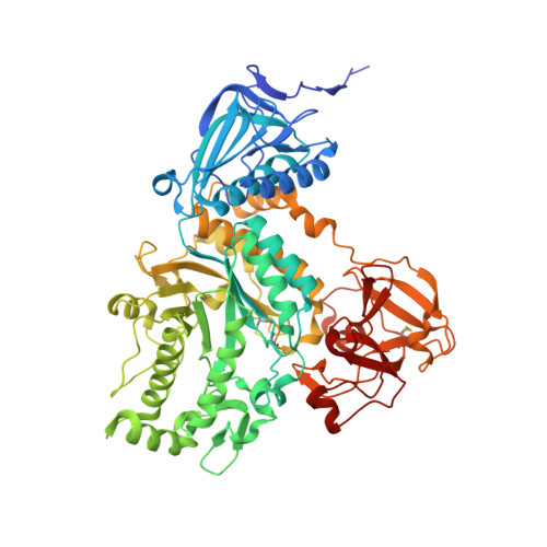
Gaining insight into the catalysis by GH20 lacto-N-biosidase using small molecule inhibitors and structural analysis

Hattie, M.Ito, T.Debowski, A.W.Arakawa, T.Katayama, T.Yamamoto, K.Fushinobu, S.Stubbs, K.A.

(2015) Chem Commun (Camb) 51: 15008-15011

  • DOI: https://doi.org/10.1039/c5cc05494j
  • Primary Citation Related Structures: 
    5BXP, 5BXR, 5BXS, 5BXT

  • PubMed Abstract: 

    The synthesis of potent inhibitors for lacto-N-biosidases and X-ray structural characterization of these compounds in complex with BbLNBase is described.


  • Organizational Affiliation
    • School of Chemistry and Biochemistry, The University of Western Australia, Crawley, WA 6009, Australia. keith.stubbs@uwa.edu.au.

Macromolecule Content 

  • Total Structure Weight: 145.03 kDa 
  • Atom Count: 10,996 
  • Modeled Residue Count: 1,270 
  • Deposited Residue Count: 1,288 
  • Unique protein chains: 1

Macromolecules

Find similar proteins by:|  3D Structure
Entity ID: 1
MoleculeChains  Sequence LengthOrganismDetailsImage
Lacto-N-biosidase
A, B
644Bifidobacterium bifidum JCM 1254Mutation(s): 0 
Gene Names: lnbB
EC: 3.2.1.140
UniProt
Find proteins for B3TLD6 (Bifidobacterium bifidum (strain DSM 20082 / JCM 1254 / BCRC 11844 / KCTC 3440 / E319f (Variant a)))
Explore B3TLD6 
Go to UniProtKB:  B3TLD6
Entity Groups
Sequence Clusters30% Identity50% Identity70% Identity90% Identity95% Identity100% Identity
UniProt GroupB3TLD6
Sequence Annotations
Expand
Reference Sequence

Small Molecules

Ligands 3 Unique
IDChains Name / Formula / InChI Key2D Diagram3D Interactions
NOK

Query on NOK


Download:Ideal Coordinates CCD File
D [auth A],
I [auth B]
2-ACETAMIDO-1,2-DIDEOXYNOJIRMYCIN
C8 H16 N2 O4
GBRAQQUMMCVTAV-LXGUWJNJSA-N
GAL

Query on GAL


Download:Ideal Coordinates CCD File
C [auth A],
H [auth B]
beta-D-galactopyranose
C6 H12 O6
WQZGKKKJIJFFOK-FPRJBGLDSA-N
SO4

Query on SO4


Download:Ideal Coordinates CCD File
E [auth A],
F [auth A],
G [auth A],
J [auth B],
K [auth B]
SULFATE ION
O4 S
QAOWNCQODCNURD-UHFFFAOYSA-L

Experimental Data & Validation

Experimental Data

  • Method: X-RAY DIFFRACTION
  • Resolution: 1.60 Å
  • R-Value Free:  0.212 (Depositor), 0.220 (DCC) 
  • R-Value Work:  0.181 (Depositor), 0.191 (DCC) 
  • R-Value Observed: 0.183 (Depositor) 
Space Group: P 21 21 2
Unit Cell:
Length ( Å )Angle ( ˚ )
a = 116.868α = 90
b = 131.271β = 90
c = 104.302γ = 90
Software Package:
Software NamePurpose
REFMACrefinement
HKL-2000data reduction
HKL-2000data scaling
REFMACphasing

Structure Validation

View Full Validation Report


Ligand Structure Quality Assessment 


Entry History 

Deposition Data

Revision History  (Full details and data files)

  • Version 1.0: 2015-09-09
    Type: Initial release
  • Version 1.1: 2015-10-14
    Changes: Database references
  • Version 1.2: 2020-02-19
    Changes: Data collection, Derived calculations
  • Version 1.3: 2020-07-29
    Type: Remediation
    Reason: Carbohydrate remediation
    Changes: Data collection, Derived calculations, Structure summary
  • Version 1.4: 2023-11-08
    Changes: Data collection, Database references, Refinement description, Structure summary
  • Version 1.5: 2024-11-13
    Changes: Structure summary