5BVW

Fragment-based discovery of potent and selective DDR1/2 inhibitors


Experimental Data Snapshot

  • Method: X-RAY DIFFRACTION
  • Resolution: 1.94 Å
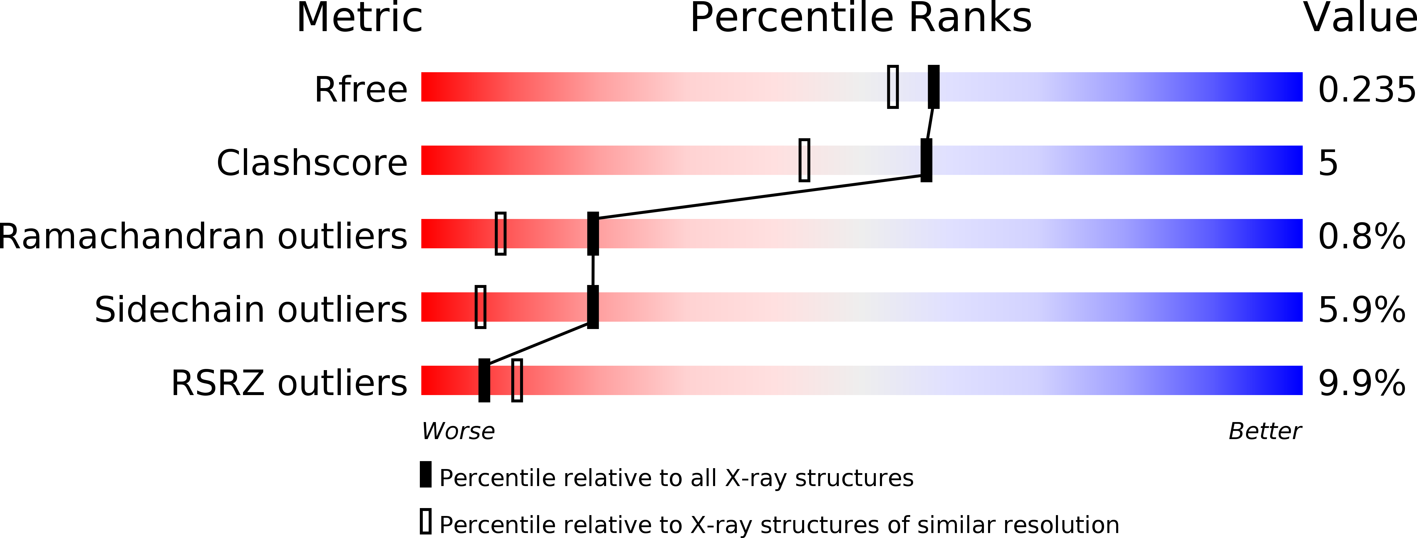
  • R-Value Free: 0.234 
  • R-Value Work: 0.184 
  • R-Value Observed: 0.186 

wwPDB Validation   3D Report Full Report


Ligand Structure Quality Assessment 

Created with Raphaël 2.3.0Worse 01 BetterLigand structure goodness of fit to experimental dataBest fitted 1N1Click on this verticalbar to view details

This is version 1.0 of the entry. See complete history


Literature

Fragment-Based Discovery of Potent and Selective DDR1/2 Inhibitors.

Murray, C.W.Berdini, V.Buck, I.M.Carr, M.E.Cleasby, A.Coyle, J.E.Curry, J.E.Day, J.E.Day, P.J.Hearn, K.Iqbal, A.Lee, L.Y.Martins, V.Mortenson, P.N.Munck, J.M.Page, L.W.Patel, S.Roomans, S.Smith, K.Tamanini, E.Saxty, G.

(2015) ACS Med Chem Lett 6: 798-803

  • DOI: https://doi.org/10.1021/acsmedchemlett.5b00143
  • Primary Citation of Related Structures:  
    5BVK, 5BVN, 5BVO, 5BVW

  • PubMed Abstract: 

    The DDR1 and DDR2 receptor tyrosine kinases are activated by extracellular collagen and have been implicated in a number of human diseases including cancer. We performed a fragment-based screen against DDR1 and identified fragments that bound either at the hinge or in the back pocket associated with the DFG-out conformation of the kinase. Modeling based on crystal structures of potent kinase inhibitors facilitated the "back-to-front" design of potent DDR1/2 inhibitors that incorporated one of the DFG-out fragments. Further optimization led to low nanomolar, orally bioavailable inhibitors that were selective for DDR1 and DDR2. The inhibitors were shown to potently inhibit DDR2 activity in cells but in contrast to unselective inhibitors such as dasatinib, they did not inhibit proliferation of mutant DDR2 lung SCC cell lines.


  • Organizational Affiliation

    Astex Pharmaceuticals, 436 Cambridge Science Park, Milton Road, Cambridge CB4 0QA, U.K.


Macromolecules
Find similar proteins by:  (by identity cutoff)  |  3D Structure
Entity ID: 1
MoleculeChains Sequence LengthOrganismDetailsImage
Epithelial discoidin domain-containing receptor 1324Homo sapiensMutation(s): 0 
Gene Names: DDR1CAKEDDR1NEPNTRK4PTK3ARTK6TRKE
EC: 2.7.10.1
UniProt & NIH Common Fund Data Resources
Find proteins for Q08345 (Homo sapiens)
Explore Q08345 
Go to UniProtKB:  Q08345
PHAROS:  Q08345
GTEx:  ENSG00000204580 
Entity Groups  
Sequence Clusters30% Identity50% Identity70% Identity90% Identity95% Identity100% Identity
UniProt GroupQ08345
Sequence Annotations
Expand
  • Reference Sequence
Small Molecules
Ligands 2 Unique
IDChains Name / Formula / InChI Key2D Diagram3D Interactions
1N1
Query on 1N1

Download Ideal Coordinates CCD File 
J [auth A]N-(2-CHLORO-6-METHYLPHENYL)-2-({6-[4-(2-HYDROXYETHYL)PIPERAZIN-1-YL]-2-METHYLPYRIMIDIN-4-YL}AMINO)-1,3-THIAZOLE-5-CARBOXAMIDE
C22 H26 Cl N7 O2 S
ZBNZXTGUTAYRHI-UHFFFAOYSA-N
IOD
Query on IOD

Download Ideal Coordinates CCD File 
B [auth A]
C [auth A]
D [auth A]
E [auth A]
F [auth A]
IODIDE ION
I
XMBWDFGMSWQBCA-UHFFFAOYSA-M
Experimental Data & Validation

Experimental Data

  • Method: X-RAY DIFFRACTION
  • Resolution: 1.94 Å
  • R-Value Free: 0.234 
  • R-Value Work: 0.184 
  • R-Value Observed: 0.186 
  • Space Group: P 21 21 21
Unit Cell:
Length ( Å )Angle ( ˚ )
a = 61.642α = 90
b = 72.78β = 90
c = 75.787γ = 90
Software Package:
Software NamePurpose
MOSFLMdata reduction
SCALAdata scaling
REFMACrefinement
PDB_EXTRACTdata extraction
Aimlessdata scaling
PHASERphasing

Structure Validation

View Full Validation Report


Ligand Structure Quality Assessment 

Created with Raphaël 2.3.0Worse 01 BetterLigand structure goodness of fit to experimental dataBest fitted 1N1Click on this verticalbar to view details

Entry History 

Revision History  (Full details and data files)

  • Version 1.0: 2015-08-05
    Type: Initial release