5BU4 | pdb_00005bu4

RIBONUCLEASE T1 COMPLEX WITH 2'GMP


Experimental Data Snapshot

  • Method: X-RAY DIFFRACTION
  • Resolution: 1.77 Å
  • R-Value Free: 
    0.225 (Depositor) 
  • R-Value Work: 
    0.172 (Depositor) 

Starting Model: experimental
View more details

wwPDB Validation 3D Report Full Report

Validation slider image for 5BU4

This is version 1.4 of the entry. See complete history

Literature

Conserved water molecules in a large family of microbial ribonucleases.

Loris, R.Langhorst, U.De Vos, S.Decanniere, K.Bouckaert, J.Maes, D.Transue, T.R.Steyaert, J.

(1999) Proteins 36: 117-134

  • DOI: https://doi.org/10.1002/(sici)1097-0134(19990701)36:1<117::aid-prot10>3.0.co;2-h
  • Primary Citation Related Structures: 
    1BU4, 2BU4, 3BU4, 4BU4, 5BU4

  • PubMed Abstract: 

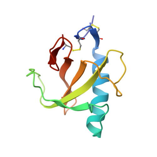
    We systematically analyzed the crystallographically determined water molecules of all known structures of RNase T1 and compared them to the ordered solvent in a large number of related microbial nucleases. To assess the crystallographers' impact on the interpretation of the solvent structure, we independently refined five validation structures from diffraction data derived from five isomorphous crystals of RNase T1. We also compared the positions of water molecules found in 11 published isomorphous RNase T1 inhibitor complexes. These data suggest that the positions of most of the waters located on the surface of a protein and that are well-determined in the experimental electron density maps are determined primarily by crystal packing forces. Water molecules with less well-defined electron density are in general unique to one or a small number of crystal structures. Only a small number of the well-defined waters are found to be independent of the crystal environment. These waters have a low accessible surface area and B-factor, and tend to be conserved in the crystal structures of a number of evolutionary related ribonucleases as well. A single water molecule is found conserved in all known microbial ribonucleases.


  • Organizational Affiliation
    • Laboratorium voor Ultrastructuur, Vlaams Interuniversitair Instituut voor Biotechnologie, Vrije Universiteit Brussel, Sint-Genesius-Rode, Belgium. reloris@vub.ac.be

Macromolecule Content 

  • Total Structure Weight: 11.5 kDa 
  • Atom Count: 903 
  • Modeled Residue Count: 104 
  • Deposited Residue Count: 104 
  • Unique protein chains: 1

Macromolecules

Find similar proteins by:|  3D Structure
Entity ID: 1
MoleculeChains  Sequence LengthOrganismDetailsImage
PROTEIN (RIBONUCLEASE T1)104Aspergillus oryzaeMutation(s): 0 
EC: 3.1.27.3 (PDB Primary Data), 4.6.1.24 (UniProt)
UniProt
Find proteins for P00651 (Aspergillus oryzae (strain ATCC 42149 / RIB 40))
Explore P00651 
Go to UniProtKB:  P00651
Entity Groups
Sequence Clusters30% Identity50% Identity70% Identity90% Identity95% Identity100% Identity
UniProt GroupP00651
Sequence Annotations
Expand
Reference Sequence

Small Molecules

Ligands 2 Unique
IDChains Name / Formula / InChI Key2D Diagram3D Interactions
2GP

Query on 2GP


Download:Ideal Coordinates CCD File
C [auth A]GUANOSINE-2'-MONOPHOSPHATE
C10 H14 N5 O8 P
WTIFIAZWCCBCGE-UUOKFMHZSA-N
CA

Query on CA


Download:Ideal Coordinates CCD File
B [auth A]CALCIUM ION
Ca
BHPQYMZQTOCNFJ-UHFFFAOYSA-N

Experimental Data & Validation

Experimental Data

  • Method: X-RAY DIFFRACTION
  • Resolution: 1.77 Å
  • R-Value Free:  0.225 (Depositor) 
  • R-Value Work:  0.172 (Depositor) 
Space Group: P 21 21 21
Unit Cell:
Length ( Å )Angle ( ˚ )
a = 40.47α = 90
b = 48.1β = 90
c = 50.78γ = 90
Software Package:
Software NamePurpose
DENZOdata reduction
SCALEPACKdata scaling
AMoREphasing
RESTRAINrefinement

Structure Validation

View Full Validation Report


Entry History 

Deposition Data

Revision History  (Full details and data files)

  • Version 1.0: 1998-09-23
    Type: Initial release
  • Version 1.1: 2007-10-16
    Changes: Version format compliance
  • Version 1.2: 2011-07-13
    Changes: Version format compliance
  • Version 1.3: 2023-09-20
    Changes: Data collection, Database references, Derived calculations, Refinement description
  • Version 1.4: 2024-11-20
    Changes: Structure summary