


Sulfolobus solfataricus P2
Christoffersen, S., Hansen, M.R., Jensen, K.S., Larsen, S., Jensen, K.F.To be published.
Experimental Data Snapshot
Entity ID: 1 | |||||
|---|---|---|---|---|---|
| Molecule | Chains | Sequence Length | Organism | Details | Image |
| Purine phosphoribosyltransferase (GpT-2) | 179 | Saccharolobus solfataricus P2 | Mutation(s): 0 Gene Names: gpT-2, SSO2424 EC: 2.4.2 | 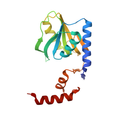 | |
UniProt | |||||
Entity Groups | |||||
| Sequence Clusters | 30% Identity50% Identity70% Identity90% Identity95% Identity100% Identity | ||||
| UniProt Group | Q97W22 | ||||
Sequence AnnotationsExpand | |||||
Reference Sequence | |||||
| Ligands 6 Unique | |||||
|---|---|---|---|---|---|
| ID | Chains | Name / Formula / InChI Key | 2D Diagram | 3D Interactions | |
| 4UO Download:Ideal Coordinates CCD File | E [auth A], J [auth B], N [auth C], S [auth D] | 2,3-dihydroxanthosine C10 H12 N4 O6 UBORTCNDUKBEOP-UUOKFMHZSA-N |  | ||
| PG4 Download:Ideal Coordinates CCD File | I [auth A], W [auth D] | TETRAETHYLENE GLYCOL C8 H18 O5 UWHCKJMYHZGTIT-UHFFFAOYSA-N |  | ||
| PGE Download:Ideal Coordinates CCD File | R [auth C] | TRIETHYLENE GLYCOL C6 H14 O4 ZIBGPFATKBEMQZ-UHFFFAOYSA-N |  | ||
| SO4 Download:Ideal Coordinates CCD File | G [auth A] H [auth A] L [auth B] P [auth C] U [auth D] | SULFATE ION O4 S QAOWNCQODCNURD-UHFFFAOYSA-L |  | ||
| PO4 Download:Ideal Coordinates CCD File | F [auth A], K [auth B], O [auth C], T [auth D] | PHOSPHATE ION O4 P NBIIXXVUZAFLBC-UHFFFAOYSA-K |  | ||
| GOL Download:Ideal Coordinates CCD File | M [auth B], Q [auth C] | GLYCEROL C3 H8 O3 PEDCQBHIVMGVHV-UHFFFAOYSA-N |  | ||
| Length ( Å ) | Angle ( ˚ ) |
|---|---|
| a = 132.218 | α = 90 |
| b = 132.218 | β = 90 |
| c = 103.102 | γ = 120 |
| Software Name | Purpose |
|---|---|
| XSCALE | data scaling |
| PHASER | phasing |
| PHENIX | refinement |
| PDB_EXTRACT | data extraction |
| Coot | model building |
| XDS | data reduction |


