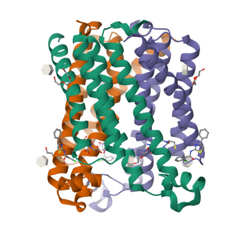
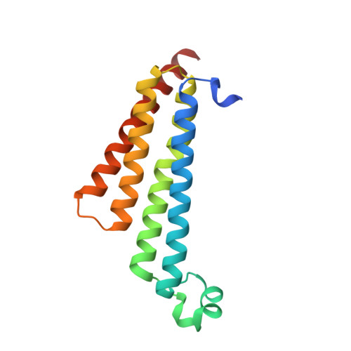
5BQG | pdb_00005bqg

Crystal Structure of mPGES-1 Bound to an Inhibitor


Experimental Data Snapshot

  • Method: X-RAY DIFFRACTION
  • Resolution: 1.44 Å
  • R-Value Free: 
    0.169 (Depositor), 0.170 (DCC) 
  • R-Value Work: 
    0.150 (Depositor), 0.150 (DCC) 
  • R-Value Observed: 
    0.151 (Depositor) 

wwPDB Validation   3D Report Full Report


Ligand Structure Quality Assessment 

Created with Raphaël 2.3.0Worse 01 BetterLigand structure goodness of fit to experimental dataBest fitted 4UJClick on this verticalbar to view detailsBest fitted GSHClick on this verticalbar to view detailsBest fitted BOGClick on this verticalbar to view detailsBest fitted JZRClick on this verticalbar to view details

This is version 1.2 of the entry. See complete history


Literature

Discovery and Characterization of 2-Acylaminoimidazole Microsomal Prostaglandin E Synthase-1 Inhibitors.

Schiffler, M.A.Antonysamy, S.Bhattachar, S.N.Campanale, K.M.Chandrasekhar, S.Condon, B.Desai, P.V.Fisher, M.J.Groshong, C.Harvey, A.Hickey, M.J.Hughes, N.E.Jones, S.A.Kim, E.J.Kuklish, S.L.Luz, J.G.Norman, B.H.Rathmell, R.E.Rizzo, J.R.Seng, T.W.Thibodeaux, S.J.Woods, T.A.York, J.S.Yu, X.P.

(2016) J Med Chem 59: 194-205

  • DOI: https://doi.org/10.1021/acs.jmedchem.5b01249
  • Primary Citation of Related Structures:  
    5BQG, 5BQH, 5BQI

  • PubMed Abstract: 

    As part of a program aimed at the discovery of antinociceptive therapy for inflammatory conditions, a screening hit was found to inhibit microsomal prostaglandin E synthase-1 (mPGES-1) with an IC50 of 17.4 μM. Structural information was used to improve enzyme potency by over 1000-fold. Addition of an appropriate substituent alleviated time-dependent cytochrome P450 3A4 (CYP3A4) inhibition. Further structure-activity relationship (SAR) studies led to 8, which had desirable potency (IC50 = 12 nM in an ex vivo human whole blood (HWB) assay) and absorption, distribution, metabolism, and excretion (ADME) properties. Studies on the formulation of 8 identified 8·H3PO4 as suitable for clinical development. Omission of a lipophilic portion of the compound led to 26, a readily orally bioavailable inhibitor with potency in HWB comparable to celecoxib. Furthermore, 26 was selective for mPGES-1 inhibition versus other mechanisms in the prostanoid pathway. These factors led to the selection of 26 as a second clinical candidate.


  • Organizational Affiliation

    Lilly Research Laboratories, A Division of Eli Lilly and Company , Indianapolis, Indiana 46285, United States.


Macromolecules
Find similar proteins by:  (by identity cutoff)  |  3D Structure
Entity ID: 1
MoleculeChains Sequence LengthOrganismDetailsImage
Prostaglandin E synthase154Homo sapiensMutation(s): 0 
Gene Names: PTGESMGST1L1MPGES1PGESPIG12
EC: 5.3.99.3 (PDB Primary Data), 1.11.1 (UniProt), 2.5.1.18 (UniProt)
Membrane Entity: Yes 
UniProt & NIH Common Fund Data Resources
Find proteins for O14684 (Homo sapiens)
Explore O14684 
Go to UniProtKB:  O14684
PHAROS:  O14684
GTEx:  ENSG00000148344 
Entity Groups  
Sequence Clusters30% Identity50% Identity70% Identity90% Identity95% Identity100% Identity
UniProt GroupO14684
Sequence Annotations
Expand
  • Reference Sequence
Small Molecules
Ligands 6 Unique
IDChains Name / Formula / InChI Key2D Diagram3D Interactions
4UJ
Query on 4UJ

Download Ideal Coordinates CCD File 
B [auth A]2-chloro-N-(4-phenyl-1,3-thiazol-2-yl)benzamide
C16 H11 Cl N2 O S
FXJFKYBQUTVGOL-UHFFFAOYSA-N
GSH
Query on GSH

Download Ideal Coordinates CCD File 
C [auth A]GLUTATHIONE
C10 H17 N3 O6 S
RWSXRVCMGQZWBV-WDSKDSINSA-N
BOG
Query on BOG

Download Ideal Coordinates CCD File 
E [auth A]octyl beta-D-glucopyranoside
C14 H28 O6
HEGSGKPQLMEBJL-RKQHYHRCSA-N
JZR
Query on JZR

Download Ideal Coordinates CCD File 
D [auth A]hexyl beta-D-glucopyranoside
C12 H24 O6
JVAZJLFFSJARQM-RMPHRYRLSA-N
PGE
Query on PGE

Download Ideal Coordinates CCD File 
F [auth A]TRIETHYLENE GLYCOL
C6 H14 O4
ZIBGPFATKBEMQZ-UHFFFAOYSA-N
PEG
Query on PEG

Download Ideal Coordinates CCD File 
G [auth A]DI(HYDROXYETHYL)ETHER
C4 H10 O3
MTHSVFCYNBDYFN-UHFFFAOYSA-N
Experimental Data & Validation

Experimental Data

  • Method: X-RAY DIFFRACTION
  • Resolution: 1.44 Å
  • R-Value Free:  0.169 (Depositor), 0.170 (DCC) 
  • R-Value Work:  0.150 (Depositor), 0.150 (DCC) 
  • R-Value Observed: 0.151 (Depositor) 
Space Group: H 3
Unit Cell:
Length ( Å )Angle ( ˚ )
a = 77.472α = 90
b = 77.472β = 90
c = 123.123γ = 120
Software Package:
Software NamePurpose
REFMACrefinement
MOSFLMdata reduction
SCALAdata scaling
PHASERphasing

Structure Validation

View Full Validation Report


Ligand Structure Quality Assessment 

Created with Raphaël 2.3.0Worse 01 BetterLigand structure goodness of fit to experimental dataBest fitted 4UJClick on this verticalbar to view detailsBest fitted GSHClick on this verticalbar to view detailsBest fitted BOGClick on this verticalbar to view detailsBest fitted JZRClick on this verticalbar to view details

Entry History 

Deposition Data

Revision History  (Full details and data files)

  • Version 1.0: 2016-04-13
    Type: Initial release
  • Version 1.1: 2020-07-29
    Type: Remediation
    Reason: Carbohydrate remediation
    Changes: Data collection, Database references, Derived calculations, Structure summary
  • Version 1.2: 2024-03-06
    Changes: Data collection, Database references, Structure summary