5BP2 | pdb_00005bp2

Dehydratase domain (DH) of a mycocerosic acid synthase-like (MAS-like) PKS, crystal form 1


Experimental Data Snapshot

  • Method: X-RAY DIFFRACTION
  • Resolution: 1.75 Å
  • R-Value Free: 
    0.203 (Depositor), 0.198 (DCC) 
  • R-Value Work: 
    0.183 (Depositor), 0.171 (DCC) 
  • R-Value Observed: 
    0.183 (Depositor) 

Starting Model: experimental
View more details

wwPDB Validation 3D Report Full Report

Validation slider image for 5BP2

This is version 1.5 of the entry. See complete history

Literature

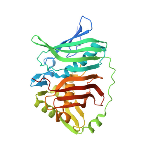
Mycocerosic acid synthase exemplifies the architecture of reducing polyketide synthases.

Herbst, D.A.Jakob, R.P.Zahringer, F.Maier, T.

(2016) Nature 531: 533-537

  • DOI: https://doi.org/10.1038/nature16993
  • Primary Citation Related Structures: 
    5BP1, 5BP2, 5BP3, 5BP4

  • PubMed Abstract: 

    Polyketide synthases (PKSs) are biosynthetic factories that produce natural products with important biological and pharmacological activities. Their exceptional product diversity is encoded in a modular architecture. Modular PKSs (modPKSs) catalyse reactions colinear to the order of modules in an assembly line, whereas iterative PKSs (iPKSs) use a single module iteratively as exemplified by fungal iPKSs (fiPKSs). However, in some cases non-colinear iterative action is also observed for modPKSs modules and is controlled by the assembly line environment. PKSs feature a structural and functional separation into a condensing and a modifying region as observed for fatty acid synthases. Despite the outstanding relevance of PKSs, the detailed organization of PKSs with complete fully reducing modifying regions remains elusive. Here we report a hybrid crystal structure of Mycobacterium smegmatis mycocerosic acid synthase based on structures of its condensing and modifying regions. Mycocerosic acid synthase is a fully reducing iPKS, closely related to modPKSs, and the prototype of mycobacterial mycocerosic acid synthase-like PKSs. It is involved in the biosynthesis of C20-C28 branched-chain fatty acids, which are important virulence factors of mycobacteria. Our structural data reveal a dimeric linker-based organization of the modifying region and visualize dynamics and conformational coupling in PKSs. On the basis of comparative small-angle X-ray scattering, the observed modifying region architecture may be common also in modPKSs. The linker-based organization provides a rationale for the characteristic variability of PKS modules as a main contributor to product diversity. The comprehensive architectural model enables functional dissection and re-engineering of PKSs.


  • Organizational Affiliation
    • Department Biozentrum, University of Basel, Klingelbergstrasse 50/70, 4056 Basel, Switzerland.

Macromolecule Content 

  • Total Structure Weight: 131.14 kDa 
  • Atom Count: 9,637 
  • Modeled Residue Count: 1,134 
  • Deposited Residue Count: 1,220 
  • Unique protein chains: 1

Macromolecules

Find similar proteins by:|  3D Structure
Entity ID: 1
MoleculeChains  Sequence LengthOrganismDetailsImage
Mycocerosic acid synthase-like polyketide synthase
A, B, C, D
305Mycolicibacterium smegmatis MC2 155Mutation(s): 0 
Gene Names: pks5MSMEG_4727
EC: 2.3.1
UniProt
Find proteins for A0R1E8 (Mycolicibacterium smegmatis (strain ATCC 700084 / mc(2)155))
Explore A0R1E8 
Go to UniProtKB:  A0R1E8
Entity Groups
Sequence Clusters30% Identity50% Identity70% Identity90% Identity95% Identity100% Identity
UniProt GroupA0R1E8
Sequence Annotations
Expand
Reference Sequence

Small Molecules

Ligands 5 Unique
IDChains Name / Formula / InChI Key2D Diagram3D Interactions
PG4

Query on PG4


Download:Ideal Coordinates CCD File
HA [auth C]TETRAETHYLENE GLYCOL
C8 H18 O5
UWHCKJMYHZGTIT-UHFFFAOYSA-N
GOL

Query on GOL


Download:Ideal Coordinates CCD File
FA [auth C],
R [auth A],
SA [auth D],
TA [auth D],
V [auth B]
GLYCEROL
C3 H8 O3
PEDCQBHIVMGVHV-UHFFFAOYSA-N
EDO

Query on EDO


Download:Ideal Coordinates CCD File
AA [auth C]
BA [auth C]
CA [auth C]
DA [auth C]
EA [auth C]
AA [auth C],
BA [auth C],
CA [auth C],
DA [auth C],
EA [auth C],
F [auth A],
G [auth A],
H [auth A],
I [auth A],
J [auth A],
JA [auth D],
K [auth A],
KA [auth D],
L [auth A],
LA [auth D],
M [auth A],
MA [auth D],
N [auth A],
NA [auth D],
O [auth A],
OA [auth D],
P [auth A],
PA [auth D],
Q [auth A],
QA [auth D],
RA [auth D],
T [auth B],
U [auth B],
W [auth C],
X [auth C],
Y [auth C],
Z [auth C]
1,2-ETHANEDIOL
C2 H6 O2
LYCAIKOWRPUZTN-UHFFFAOYSA-N
CL

Query on CL


Download:Ideal Coordinates CCD File
GA [auth C],
S [auth A],
UA [auth D]
CHLORIDE ION
Cl
VEXZGXHMUGYJMC-UHFFFAOYSA-M
MG

Query on MG


Download:Ideal Coordinates CCD File
E [auth A],
IA [auth D]
MAGNESIUM ION
Mg
JLVVSXFLKOJNIY-UHFFFAOYSA-N

Experimental Data & Validation

Experimental Data

  • Method: X-RAY DIFFRACTION
  • Resolution: 1.75 Å
  • R-Value Free:  0.203 (Depositor), 0.198 (DCC) 
  • R-Value Work:  0.183 (Depositor), 0.171 (DCC) 
  • R-Value Observed: 0.183 (Depositor) 
Space Group: P 1 21 1
Unit Cell:
Length ( Å )Angle ( ˚ )
a = 59.65α = 90
b = 162.4β = 91.38
c = 66.62γ = 90
Software Package:
Software NamePurpose
BUSTERrefinement
XDSdata reduction
XSCALEdata scaling
PHASERphasing

Structure Validation

View Full Validation Report


Entry History 

& Funding Information

Deposition Data


Funding OrganizationLocationGrant Number
Swiss National Science FoundationSwitzerland125357
Swiss National Science FoundationSwitzerland138262
Swiss National Science FoundationSwitzerland159696
Swiss National Science FoundationSwitzerland145023

Revision History  (Full details and data files)

  • Version 1.0: 2016-03-09
    Type: Initial release
  • Version 1.1: 2016-03-30
    Changes: Database references
  • Version 1.2: 2016-04-06
    Changes: Database references
  • Version 1.3: 2016-12-28
    Changes: Structure summary
  • Version 1.4: 2017-09-06
    Changes: Author supporting evidence
  • Version 1.5: 2024-01-10
    Changes: Data collection, Database references, Derived calculations, Refinement description