5BOB | pdb_00005bob

Crystal Structure of the Meningitis Pathogen Streptococcus suis adhesion Fhb


Experimental Data Snapshot

  • Method: X-RAY DIFFRACTION
  • Resolution: 1.50 Å
  • R-Value Free: 
    0.189 (Depositor), 0.189 (DCC) 
  • R-Value Work: 
    0.161 (Depositor), 0.161 (DCC) 
  • R-Value Observed: 
    0.163 (Depositor) 

wwPDB Validation 3D Report Full Report

Validation slider image for 5BOB

This is version 1.1 of the entry. See complete history

Literature

Expression, purification, crystallization and structure determination of the N terminal domain of Fhb, a factor H binding protein from Streptococcus suis.

Zhang, C.Yu, Y.Yang, M.Jiang, Y.

(2015) Biochem Biophys Res Commun 466: 413-417

  • DOI: https://doi.org/10.1016/j.bbrc.2015.09.040
  • Primary Citation Related Structures: 
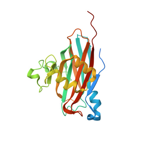
    5BOB

  • PubMed Abstract: 

    Fhb is a surface virulence protein from Streptococcus suis, which could aid bacterial evasion of host innate immune defense by recruiting complement regulator factor H to inactivate C3b deposited on bacterial surface in blood. Here we successfully expressed and purified the N terminal domain of Fhb (N-Fhb) and obtained crystals of the N-Fhb by sitting-drop vapor diffusion method with a resolution of 1.50 Å. The crystals belong to space group C2 with unit cell parameters a = 127.1 Å, b = 77.3 Å, c = 131.6 Å, α = 90°, β = 115.9°, γ = 90°. The structure of N-Fhb was determined by SAD method and the core structure of N-Fhb is a β sandwich. We speculated that binding of Fhb to human factor H may be mainly mediated by surface amino acids with negative charges.


  • Organizational Affiliation
    • State Key Laboratory of Pathogen and Biosecurity, Beijng Institute of Microbiology and Infectious Disease, No. 20 Dongda Street, Fengtai District, Beijing 100071, China.

Macromolecule Content 

  • Total Structure Weight: 129.64 kDa 
  • Atom Count: 9,071 
  • Modeled Residue Count: 996 
  • Deposited Residue Count: 1,130 
  • Unique protein chains: 1

Macromolecules

Find similar proteins by:|  3D Structure
Entity ID: 1
MoleculeChains  Sequence LengthOrganismDetailsImage
Translation initiation factor 2 (IF-2 GTPase)
A, B, C, D, E
226Streptococcus suis 05ZYH33Mutation(s): 0 
Gene Names: SSU05_0272

Small Molecules

Ligands 1 Unique
IDChains Name / Formula / InChI Key2D Diagram3D Interactions
GOL

Query on GOL


Download:Ideal Coordinates CCD File
F [auth A]
G [auth A]
H [auth B]
I [auth B]
J [auth B]
F [auth A],
G [auth A],
H [auth B],
I [auth B],
J [auth B],
K [auth C],
L [auth D],
M [auth D],
N [auth E],
O [auth E],
P [auth E]
GLYCEROL
C3 H8 O3
PEDCQBHIVMGVHV-UHFFFAOYSA-N
Modified Residues  1 Unique
IDChains TypeFormula2D DiagramParent
MSE
Query on MSE
A, B, C, D, E
L-PEPTIDE LINKINGC5 H11 N O2 SeMET

Experimental Data & Validation

Experimental Data

  • Method: X-RAY DIFFRACTION
  • Resolution: 1.50 Å
  • R-Value Free:  0.189 (Depositor), 0.189 (DCC) 
  • R-Value Work:  0.161 (Depositor), 0.161 (DCC) 
  • R-Value Observed: 0.163 (Depositor) 
Space Group: C 1 2 1
Unit Cell:
Length ( Å )Angle ( ˚ )
a = 127.075α = 90
b = 77.341β = 115.93
c = 131.642γ = 90
Software Package:
Software NamePurpose
REFMACrefinement
SCALEPACKdata scaling
PDB_EXTRACTdata extraction
HKL-2000data reduction
PHASERphasing

Structure Validation

View Full Validation Report


Entry History 

Deposition Data

Revision History  (Full details and data files)

  • Version 1.0: 2015-11-18
    Type: Initial release
  • Version 1.1: 2024-11-13
    Changes: Data collection, Database references, Derived calculations, Structure summary