5BN3

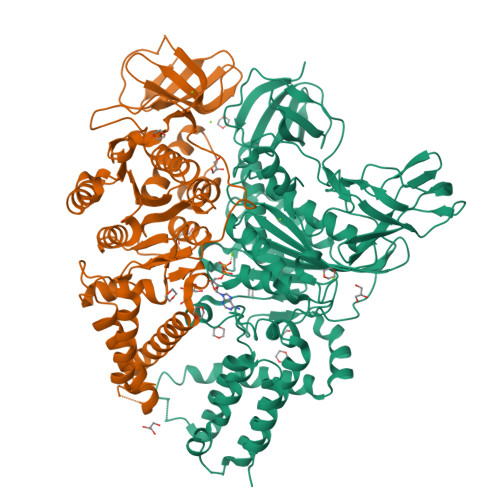
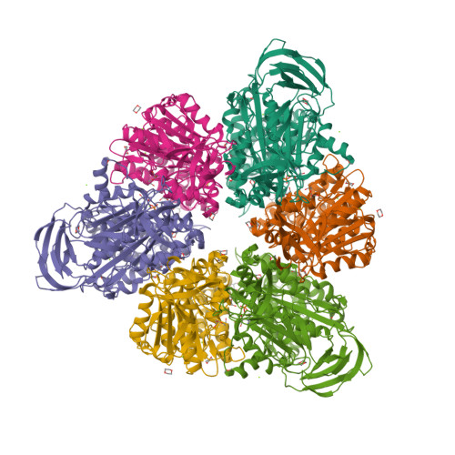
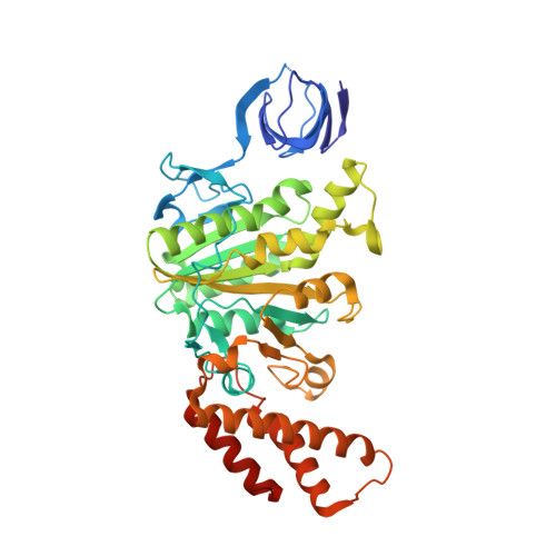
Structure of a unique ATP synthase NeqA-NeqB in complex with ADP from Nanoarcheaum equitans


Experimental Data Snapshot

  • Method: X-RAY DIFFRACTION
  • Resolution: 2.00 Å
  • R-Value Free: 0.210 
  • R-Value Work: 0.184 
  • R-Value Observed: 0.185 

Starting Model: experimental
View more details

wwPDB Validation   3D Report Full Report


Ligand Structure Quality Assessment 


This is version 1.3 of the entry. See complete history


Literature

Structural Basis for a Unique ATP Synthase Core Complex from Nanoarcheaum equitans

Mohanty, S.Jobichen, C.Chichili, V.P.R.Velazquez-Campoy, A.Low, B.C.Hogue, C.W.V.Sivaraman, J.

(2015) J Biol Chem 290: 27280-27296

  • DOI: https://doi.org/10.1074/jbc.M115.677492
  • Primary Citation of Related Structures:  
    5BN3, 5BN4, 5BN5, 5BO5

  • PubMed Abstract: 

    ATP synthesis is a critical and universal life process carried out by ATP synthases. Whereas eukaryotic and prokaryotic ATP synthases are well characterized, archaeal ATP synthases are relatively poorly understood. The hyperthermophilic archaeal parasite, Nanoarcheaum equitans, lacks several subunits of the ATP synthase and is suspected to be energetically dependent on its host, Ignicoccus hospitalis. This suggests that this ATP synthase might be a rudimentary machine. Here, we report the crystal structures and biophysical studies of the regulatory subunit, NeqB, the apo-NeqAB, and NeqAB in complex with nucleotides, ADP, and adenylyl-imidodiphosphate (non-hydrolysable analog of ATP). NeqB is ∼20 amino acids shorter at its C terminus than its homologs, but this does not impede its binding with NeqA to form the complex. The heterodimeric NeqAB complex assumes a closed, rigid conformation irrespective of nucleotide binding; this differs from its homologs, which require conformational changes for catalytic activity. Thus, although N. equitans possesses an ATP synthase core A3B3 hexameric complex, it might not function as a bona fide ATP synthase.


  • Organizational Affiliation

    Department of Biological Sciences, National University of Singapore, Singapore 117543, Singapore.


Macromolecules
Find similar proteins by:  (by identity cutoff)  |  3D Structure
Entity ID: 1
MoleculeChains Sequence LengthOrganismDetailsImage
V-type ATP synthase alpha chain570Nanoarchaeum equitans Kin4-MMutation(s): 0 
Gene Names: atpANEQ103
EC: 3.6.3.14 (PDB Primary Data), 7.1.2.2 (UniProt)
UniProt
Find proteins for Q74MJ7 (Nanoarchaeum equitans (strain Kin4-M))
Explore Q74MJ7 
Go to UniProtKB:  Q74MJ7
Entity Groups  
Sequence Clusters30% Identity50% Identity70% Identity90% Identity95% Identity100% Identity
UniProt GroupQ74MJ7
Sequence Annotations
Expand
  • Reference Sequence
Find similar proteins by:  (by identity cutoff)  |  3D Structure
Entity ID: 2
MoleculeChains Sequence LengthOrganismDetailsImage
NEQ263416Nanoarchaeum equitans Kin4-MMutation(s): 0 
Gene Names: NEQ263
UniProt
Find proteins for Q74MS5 (Nanoarchaeum equitans (strain Kin4-M))
Explore Q74MS5 
Go to UniProtKB:  Q74MS5
Entity Groups  
Sequence Clusters30% Identity50% Identity70% Identity90% Identity95% Identity100% Identity
UniProt GroupQ74MS5
Sequence Annotations
Expand
  • Reference Sequence
Small Molecules
Ligands 5 Unique
IDChains Name / Formula / InChI Key2D Diagram3D Interactions
ADP
Query on ADP

Download Ideal Coordinates CCD File 
C [auth A]ADENOSINE-5'-DIPHOSPHATE
C10 H15 N5 O10 P2
XTWYTFMLZFPYCI-KQYNXXCUSA-N
SO4
Query on SO4

Download Ideal Coordinates CCD File 
X [auth B]SULFATE ION
O4 S
QAOWNCQODCNURD-UHFFFAOYSA-L
GOL
Query on GOL

Download Ideal Coordinates CCD File 
H [auth A]
I [auth A]
J [auth A]
Q [auth B]
R [auth B]
H [auth A],
I [auth A],
J [auth A],
Q [auth B],
R [auth B],
S [auth B],
T [auth B]
GLYCEROL
C3 H8 O3
PEDCQBHIVMGVHV-UHFFFAOYSA-N
DIO
Query on DIO

Download Ideal Coordinates CCD File 
D [auth A]
E [auth A]
F [auth A]
G [auth A]
M [auth B]
D [auth A],
E [auth A],
F [auth A],
G [auth A],
M [auth B],
N [auth B],
O [auth B],
P [auth B]
1,4-DIETHYLENE DIOXIDE
C4 H8 O2
RYHBNJHYFVUHQT-UHFFFAOYSA-N
MG
Query on MG

Download Ideal Coordinates CCD File 
K [auth A],
L [auth A],
U [auth B],
V [auth B],
W [auth B]
MAGNESIUM ION
Mg
JLVVSXFLKOJNIY-UHFFFAOYSA-N
Experimental Data & Validation

Experimental Data

  • Method: X-RAY DIFFRACTION
  • Resolution: 2.00 Å
  • R-Value Free: 0.210 
  • R-Value Work: 0.184 
  • R-Value Observed: 0.185 
  • Space Group: H 3
Unit Cell:
Length ( Å )Angle ( ˚ )
a = 192.459α = 90
b = 192.459β = 90
c = 110.241γ = 120
Software Package:
Software NamePurpose
PHENIXrefinement
HKL-2000data reduction
HKL-2000data scaling
PHENIXphasing

Structure Validation

View Full Validation Report


Ligand Structure Quality Assessment 


Entry History 

Deposition Data

Revision History  (Full details and data files)

  • Version 1.0: 2015-09-16
    Type: Initial release
  • Version 1.1: 2015-10-14
    Changes: Database references
  • Version 1.2: 2015-11-18
    Changes: Database references
  • Version 1.3: 2023-11-08
    Changes: Data collection, Database references, Derived calculations, Refinement description