


X-ray Crystal Structure of a 2-C-methyl-D-erythritol 2,4-cyclodiphosphate synthase from Pseudomonas aeruginosa
SSGCID, Fairman, J.W., Dranow, D.M., Lorimer, D., Edwards, T.E.To be published.
Experimental Data Snapshot
Starting Model: experimental
View more details
Entity ID: 1 | |||||
|---|---|---|---|---|---|
| Molecule | Chains | Sequence Length | Organism | Details | Image |
| 2-C-methyl-D-erythritol 2,4-cyclodiphosphate synthase | 164 | Pseudomonas aeruginosa PAO1 | Mutation(s): 0 Gene Names: ispF, PA3627 EC: 4.6.1.12 | 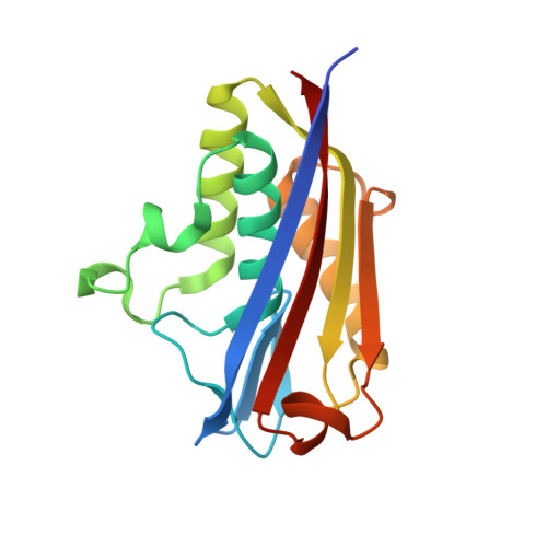 | |
UniProt | |||||
Entity Groups | |||||
| Sequence Clusters | 30% Identity50% Identity70% Identity90% Identity95% Identity100% Identity | ||||
| UniProt Group | P57708 | ||||
Sequence AnnotationsExpand | |||||
Reference Sequence | |||||
| Ligands 4 Unique | |||||
|---|---|---|---|---|---|
| ID | Chains | Name / Formula / InChI Key | 2D Diagram | 3D Interactions | |
| C5P Download:Ideal Coordinates CCD File | E [auth A], H [auth B], N [auth C] | CYTIDINE-5'-MONOPHOSPHATE C9 H14 N3 O8 P IERHLVCPSMICTF-XVFCMESISA-N |  | ||
| MPD Download:Ideal Coordinates CCD File | F [auth A] | (4S)-2-METHYL-2,4-PENTANEDIOL C6 H14 O2 SVTBMSDMJJWYQN-YFKPBYRVSA-N |  | ||
| PO4 Download:Ideal Coordinates CCD File | I [auth B], J [auth B], K [auth B], L [auth B] | PHOSPHATE ION O4 P NBIIXXVUZAFLBC-UHFFFAOYSA-K |  | ||
| MG Download:Ideal Coordinates CCD File | D [auth A], G [auth B], M [auth C] | MAGNESIUM ION Mg JLVVSXFLKOJNIY-UHFFFAOYSA-N |  | ||
| Length ( Å ) | Angle ( ˚ ) |
|---|---|
| a = 80.56 | α = 90 |
| b = 100.21 | β = 90 |
| c = 54.81 | γ = 90 |
| Software Name | Purpose |
|---|---|
| XSCALE | data scaling |
| PHENIX | refinement |
| PDB_EXTRACT | data extraction |
| XDS | data processing |
| XDS | data reduction |
| BALBES | phasing |
| Funding Organization | Location | Grant Number |
|---|---|---|
| National Institutes of Health/National Institute Of Allergy and Infectious Diseases (NIH/NIAID) | United States | -- |


