5B5U | pdb_00005b5u

Crystal structure of truncated Pyrococcus furiosus L-asparaginase with peptide


Experimental Data Snapshot

  • Method: X-RAY DIFFRACTION
  • Resolution: 2.61 Å
  • R-Value Free: 
    0.242 (Depositor), 0.234 (DCC) 
  • R-Value Work: 
    0.203 (Depositor), 0.202 (DCC) 
  • R-Value Observed: 
    0.205 (Depositor) 

Starting Model: experimental
View more details

wwPDB Validation 3D Report Full Report

Validation slider image for 5B5U

This is version 1.3 of the entry. See complete history

Literature

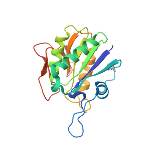
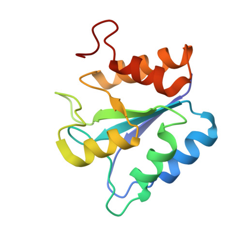
Heat induces end to end repetitive association in P. furiosus L-asparaginase which enables its thermophilic property.

Sharma, P.Tomar, R.Yadav, S.S.Badmalia, M.D.Nath, S.K.Kundu, B.

(2020) Sci Rep 10: 21702-21702

  • DOI: https://doi.org/10.1038/s41598-020-78877-z
  • Primary Citation Related Structures: 
    5B5U, 5B74, 5CBP

  • PubMed Abstract: 

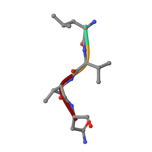
    It remains undeciphered how thermophilic enzymes display enhanced stability at elevated temperatures. Taking L-asparaginase from P. furiosus (PfA) as an example, we combined scattering shapes deduced from small-angle X-ray scattering (SAXS) data at increased temperatures with symmetry mates from crystallographic structures to find that heating caused end-to-end association. The small contact point of self-binding appeared to be enabled by a terminal short β-strand in N-terminal domain, Leu 179 -Val-Val-Asn 182 (LVVN). Interestingly, deletion of this strand led to a defunct enzyme, whereas suplementation of the peptide LVVN to the defunct enzyme restored structural frameworkwith mesophile-type functionality. Crystal structure of the peptide-bound defunct enzyme showed that one peptide ispresent in the same coordinates as in original enzyme, explaining gain-of lost function. A second peptide was seen bound to the protein at a different location suggesting its possible role in substrate-free molecular-association. Overall, we show that the heating induced self-assembly of native shapes of PfA led to an apparent super-stable assembly.


  • Organizational Affiliation
    • CSIR-Institute of Microbial Technology, Sec 39 A, Chandigarh, 160036, India.

Macromolecule Content 

  • Total Structure Weight: 34.86 kDa 
  • Atom Count: 2,436 
  • Modeled Residue Count: 310 
  • Deposited Residue Count: 313 
  • Unique protein chains: 3

Macromolecules

Find similar proteins by:|  3D Structure
Entity ID: 1
MoleculeChains  Sequence LengthOrganismDetailsImage
L-asparaginase178Pyrococcus furiosus DSM 3638Mutation(s): 0 
Gene Names: PF2047
EC: 3.5.1.1
UniProt
Find proteins for Q8TZE8 (Pyrococcus furiosus (strain ATCC 43587 / DSM 3638 / JCM 8422 / Vc1))
Explore Q8TZE8 
Go to UniProtKB:  Q8TZE8
Entity Groups
Sequence Clusters30% Identity50% Identity70% Identity90% Identity95% Identity100% Identity
UniProt GroupQ8TZE8
Sequence Annotations
Expand
Reference Sequence
Find similar proteins by:|  3D Structure
Entity ID: 2
MoleculeChains  Sequence LengthOrganismDetailsImage
L-asparaginase127Pyrococcus furiosus DSM 3638Mutation(s): 0 
Gene Names: PF2047
EC: 3.5.1.1
UniProt
Find proteins for Q8TZE8 (Pyrococcus furiosus (strain ATCC 43587 / DSM 3638 / JCM 8422 / Vc1))
Explore Q8TZE8 
Go to UniProtKB:  Q8TZE8
Entity Groups
Sequence Clusters30% Identity50% Identity70% Identity90% Identity95% Identity100% Identity
UniProt GroupQ8TZE8
Sequence Annotations
Expand
Reference Sequence
Find similar proteins by:  Sequence   |   3D Structure  
Entity ID: 3
MoleculeChains  Sequence LengthOrganismDetailsImage
LEU-VAL-VAL-ASNC,
D [auth E]
4Pyrococcus furiosusMutation(s): 0 

Small Molecules

Ligands 4 Unique
IDChains Name / Formula / InChI Key2D Diagram3D Interactions
ASP

Query on ASP


Download:Ideal Coordinates CCD File
E [auth A]ASPARTIC ACID
C4 H7 N O4
CKLJMWTZIZZHCS-REOHCLBHSA-N
GOL

Query on GOL


Download:Ideal Coordinates CCD File
I [auth A],
J [auth A],
K [auth A]
GLYCEROL
C3 H8 O3
PEDCQBHIVMGVHV-UHFFFAOYSA-N
IPA

Query on IPA


Download:Ideal Coordinates CCD File
G [auth A],
H [auth A]
ISOPROPYL ALCOHOL
C3 H8 O
KFZMGEQAYNKOFK-UHFFFAOYSA-N
CL

Query on CL


Download:Ideal Coordinates CCD File
F [auth A],
L [auth B]
CHLORIDE ION
Cl
VEXZGXHMUGYJMC-UHFFFAOYSA-M

Experimental Data & Validation

Experimental Data

  • Method: X-RAY DIFFRACTION
  • Resolution: 2.61 Å
  • R-Value Free:  0.242 (Depositor), 0.234 (DCC) 
  • R-Value Work:  0.203 (Depositor), 0.202 (DCC) 
  • R-Value Observed: 0.205 (Depositor) 
Space Group: P 65 2 2
Unit Cell:
Length ( Å )Angle ( ˚ )
a = 91.31α = 90
b = 91.31β = 90
c = 186.971γ = 120
Software Package:
Software NamePurpose
PHENIXrefinement
iMOSFLMdata reduction
SCALAdata scaling
MOLREPphasing

Structure Validation

View Full Validation Report


Entry History 

Deposition Data

Revision History  (Full details and data files)

  • Version 1.0: 2017-05-31
    Type: Initial release
  • Version 1.1: 2022-06-08
    Changes: Database references, Structure summary
  • Version 1.2: 2023-11-08
    Changes: Data collection, Refinement description
  • Version 1.3: 2024-11-13
    Changes: Structure summary