5B1H | pdb_00005b1h

Crystal structure of cystathionine beta-synthase from Lactobacillus plantarum


Experimental Data Snapshot

  • Method: X-RAY DIFFRACTION
  • Resolution: 2.40 Å
  • R-Value Free: 
    0.221 (Depositor), 0.223 (DCC) 
  • R-Value Work: 
    0.189 (Depositor), 0.190 (DCC) 
  • R-Value Observed: 
    0.189 (Depositor) 

Starting Model: experimental
View more details

wwPDB Validation 3D Report Full Report

Validation slider image for 5B1H

This is version 1.5 of the entry. See complete history

Literature

Crystallographic and mutational analyses of cystathionine beta-synthase in the H2 S-synthetic gene cluster in Lactobacillus plantarum

Matoba, Y.Yoshida, T.Izuhara-Kihara, H.Noda, M.Sugiyama, M.

(2017) Protein Sci 26: 763-783

  • DOI: https://doi.org/10.1002/pro.3123
  • Primary Citation Related Structures: 
    5B1H, 5B1I

  • PubMed Abstract: 

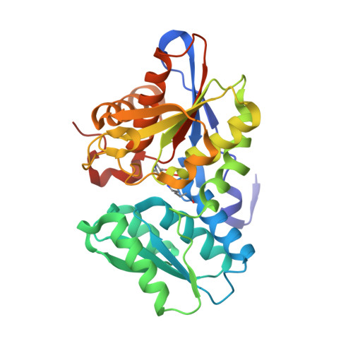
    Cystathionine β-synthase (CBS) catalyzes the formation of l-cystathionine from l-serine and l-homocysteine. The resulting l-cystathionine is decomposed into l-cysteine, ammonia, and α-ketobutylic acid by cystathionine γ-lyase (CGL). This reverse transsulfuration pathway, which is catalyzed by both enzymes, mainly occurs in eukaryotic cells. The eukaryotic CBS and CGL have recently been recognized as major physiological enzymes for the generation of hydrogen sulfide (H 2 S). In some bacteria, including the plant-derived lactic acid bacterium Lactobacillus plantarum, the CBS- and CGL-encoding genes form a cluster in their genomes. Inactivation of these enzymes has been reported to suppress H 2 S production in bacteria; interestingly, it has been shown that H 2 S suppression increases their susceptibility to various antibiotics. In the present study, we characterized the enzymatic properties of the L. plantarum CBS, whose amino acid sequence displays a similarity with those of O-acetyl-l-serine sulfhydrylase (OASS) that catalyzes the generation of l-cysteine from O-acetyl-l-serine (l-OAS) and H 2 S. The L. plantarum CBS shows l-OAS- and l-cysteine-dependent CBS activities together with OASS activity. Especially, it catalyzes the formation of H 2 S in the presence of l-cysteine and l-homocysteine, together with the formation of l-cystathionine. The high affinity toward l-cysteine as a first substrate and tendency to use l-homocysteine as a second substrate might be associated with its enzymatic ability to generate H 2 S. Crystallographic and mutational analyses of CBS indicate that the Ala70 and Glu223 residues at the substrate binding pocket are important for the H 2 S-generating activity.


  • Organizational Affiliation
    • Graduate School of Biomedical and Health Sciences, Hiroshima University, Kasumi 1-2-3, Minami-ku, Hiroshima, 734-8551, Japan.

Macromolecule Content 

  • Total Structure Weight: 136.59 kDa 
  • Atom Count: 9,736 
  • Modeled Residue Count: 1,212 
  • Deposited Residue Count: 1,244 
  • Unique protein chains: 1

Macromolecules

Find similar proteins by:|  3D Structure
Entity ID: 1
MoleculeChains  Sequence LengthOrganismDetailsImage
Cystathionine beta-synthase
A, B, C, D
311Lactiplantibacillus plantarum WCFS1Mutation(s): 0 
Gene Names: cbslp_0256
EC: 4.2.1.22 (PDB Primary Data), 2.5.1.47 (UniProt)
UniProt
Find proteins for F9UT54 (Lactiplantibacillus plantarum (strain ATCC BAA-793 / NCIMB 8826 / WCFS1))
Explore F9UT54 
Go to UniProtKB:  F9UT54
Entity Groups
Sequence Clusters30% Identity50% Identity70% Identity90% Identity95% Identity100% Identity
UniProt GroupF9UT54
Sequence Annotations
Expand
Reference Sequence

Small Molecules

Ligands 2 Unique
IDChains Name / Formula / InChI Key2D Diagram3D Interactions
SO4

Query on SO4


Download:Ideal Coordinates CCD File
AA [auth D]
E [auth A]
F [auth A]
G [auth A]
H [auth A]
AA [auth D],
E [auth A],
F [auth A],
G [auth A],
H [auth A],
M [auth B],
N [auth B],
S [auth C],
T [auth C],
U [auth C],
V [auth C],
Z [auth D]
SULFATE ION
O4 S
QAOWNCQODCNURD-UHFFFAOYSA-L
GOL

Query on GOL


Download:Ideal Coordinates CCD File
BA [auth D]
CA [auth D]
DA [auth D]
I [auth A]
J [auth A]
BA [auth D],
CA [auth D],
DA [auth D],
I [auth A],
J [auth A],
K [auth A],
L [auth A],
O [auth B],
P [auth B],
Q [auth B],
R [auth B],
W [auth C],
X [auth C],
Y [auth C]
GLYCEROL
C3 H8 O3
PEDCQBHIVMGVHV-UHFFFAOYSA-N
Modified Residues  1 Unique
IDChains TypeFormula2D DiagramParent
LLP
Query on LLP
A, B, C, D
L-PEPTIDE LINKINGC14 H22 N3 O7 PLYS

Experimental Data & Validation

Experimental Data

  • Method: X-RAY DIFFRACTION
  • Resolution: 2.40 Å
  • R-Value Free:  0.221 (Depositor), 0.223 (DCC) 
  • R-Value Work:  0.189 (Depositor), 0.190 (DCC) 
  • R-Value Observed: 0.189 (Depositor) 
Space Group: C 1 2 1
Unit Cell:
Length ( Å )Angle ( ˚ )
a = 139.89α = 90
b = 146.34β = 90
c = 82.79γ = 90
Software Package:
Software NamePurpose
CNSrefinement
AMoREdata reduction
SCALAdata scaling
MOLREPphasing

Structure Validation

View Full Validation Report


Entry History 

Deposition Data

Revision History  (Full details and data files)

  • Version 1.0: 2016-12-07
    Type: Initial release
  • Version 1.1: 2017-02-15
    Changes: Database references
  • Version 1.2: 2017-04-05
    Changes: Database references
  • Version 1.3: 2020-02-26
    Changes: Data collection
  • Version 1.4: 2023-11-08
    Changes: Data collection, Database references, Refinement description
  • Version 1.5: 2023-11-15
    Changes: Data collection