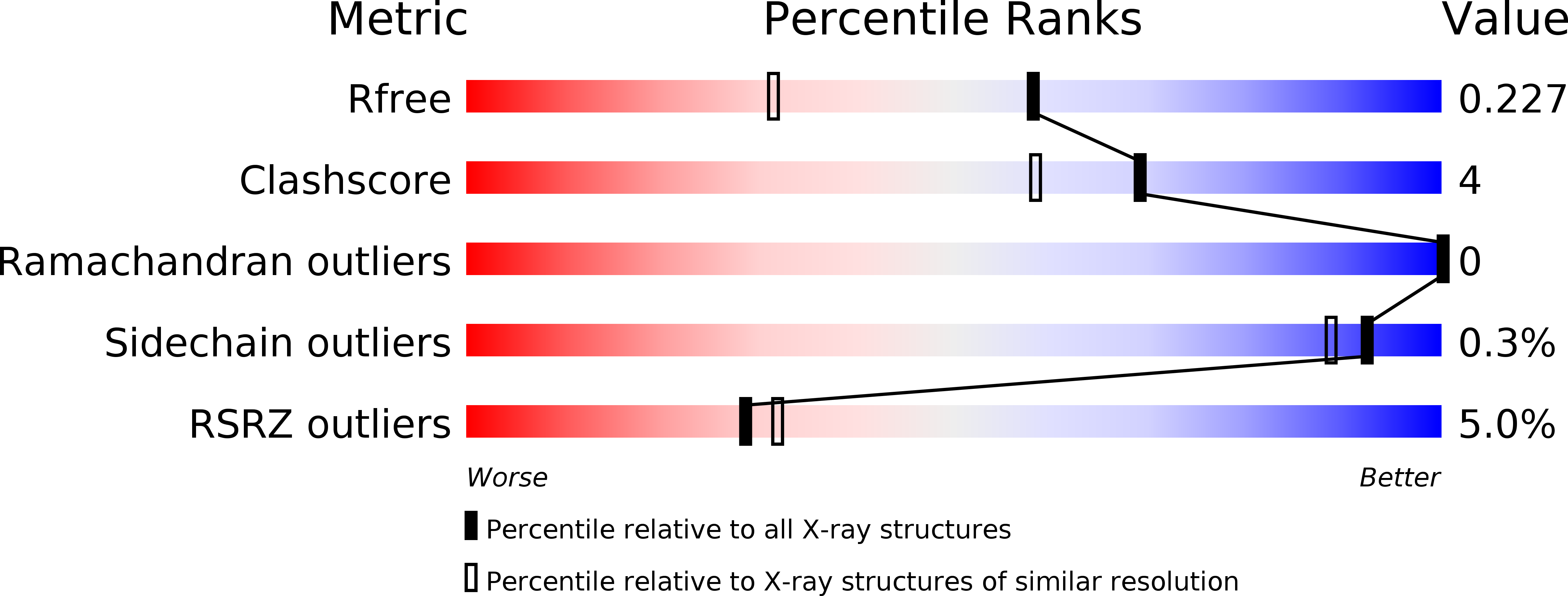
5B0U

Crystal structure of the mutated 19 kDa protein of Oplophorus luciferase (nanoKAZ)


Experimental Data Snapshot

  • Method: X-RAY DIFFRACTION
  • Resolution: 1.71 Å
  • R-Value Free: 0.226 
  • R-Value Work: 0.201 
  • R-Value Observed: 0.202 

wwPDB Validation   3D Report Full Report


This is version 1.4 of the entry. See complete history


Literature

Crystal structure of nanoKAZ: The mutated 19 kDa component of Oplophorus luciferase catalyzing the bioluminescent reaction with coelenterazine

Tomabechi, Y.Hosoya, T.Ehara, H.Sekine, S.Shirouzu, M.Inouye, S.

(2016) Biochem Biophys Res Commun 470: 88-93

  • DOI: https://doi.org/10.1016/j.bbrc.2015.12.123
  • Primary Citation of Related Structures:  
    5B0U

  • PubMed Abstract: 

    The 19 kDa protein (KAZ) of Oplophorus luciferase is a catalytic component, that oxidizes coelenterazine (a luciferin) with molecular oxygen to emit light. The crystal structure of the mutated 19 kDa protein (nanoKAZ) was determined at 1.71 Å resolution. The structure consists of 11 antiparallel β-strands forming a β-barrel that is capped by 4 short α-helices. The structure of nanoKAZ is similar to those of fatty acid-binding proteins (FABPs), even though the amino acid sequence similarity was very low between them. The coelenterazine-binding site and the catalytic site for the luminescence reaction might be in a central cavity of the β-barrel structure.


  • Organizational Affiliation

    Division of Structural and Synthetic Biology, RIKEN Center for Life Science Technologies, 1-7-22 Suehiro-cho, Tsurumi-ku, Yokohama 230-0045, Japan.


Macromolecules
Find similar proteins by:  (by identity cutoff)  |  3D Structure
Entity ID: 1
MoleculeChains Sequence LengthOrganismDetailsImage
Oplophorus-luciferin 2-monooxygenase catalytic subunit
A, B
191Oplophorus gracilirostrisMutation(s): 16 
EC: 1.13.12.13
UniProt
Find proteins for Q9GV45 (Oplophorus gracilirostris)
Explore Q9GV45 
Go to UniProtKB:  Q9GV45
Entity Groups  
Sequence Clusters30% Identity50% Identity70% Identity90% Identity95% Identity100% Identity
UniProt GroupQ9GV45
Sequence Annotations
Expand
  • Reference Sequence
Experimental Data & Validation

Experimental Data

  • Method: X-RAY DIFFRACTION
  • Resolution: 1.71 Å
  • R-Value Free: 0.226 
  • R-Value Work: 0.201 
  • R-Value Observed: 0.202 
  • Space Group: P 42 21 2
Unit Cell:
Length ( Å )Angle ( ˚ )
a = 79.067α = 90
b = 79.067β = 90
c = 123.425γ = 90
Software Package:
Software NamePurpose
HKL-2000data collection
PHASERphasing
PHENIXrefinement
PDB_EXTRACTdata extraction
HKL-2000data reduction
HKL-2000data scaling

Structure Validation

View Full Validation Report


Entry History 

Deposition Data

Revision History  (Full details and data files)

  • Version 1.0: 2016-01-13
    Type: Initial release
  • Version 1.1: 2016-01-27
    Changes: Database references
  • Version 1.2: 2016-02-10
    Changes: Database references
  • Version 1.3: 2020-02-26
    Changes: Data collection, Database references, Derived calculations
  • Version 1.4: 2024-03-20
    Changes: Data collection, Database references