5AZ1

Crystal structure of aldo-keto reductase (AKR2E5) complexed with NADPH


Experimental Data Snapshot

  • Method: X-RAY DIFFRACTION
  • Resolution: 2.30 Å
  • R-Value Free: 0.206 
  • R-Value Work: 0.159 
  • R-Value Observed: 0.161 

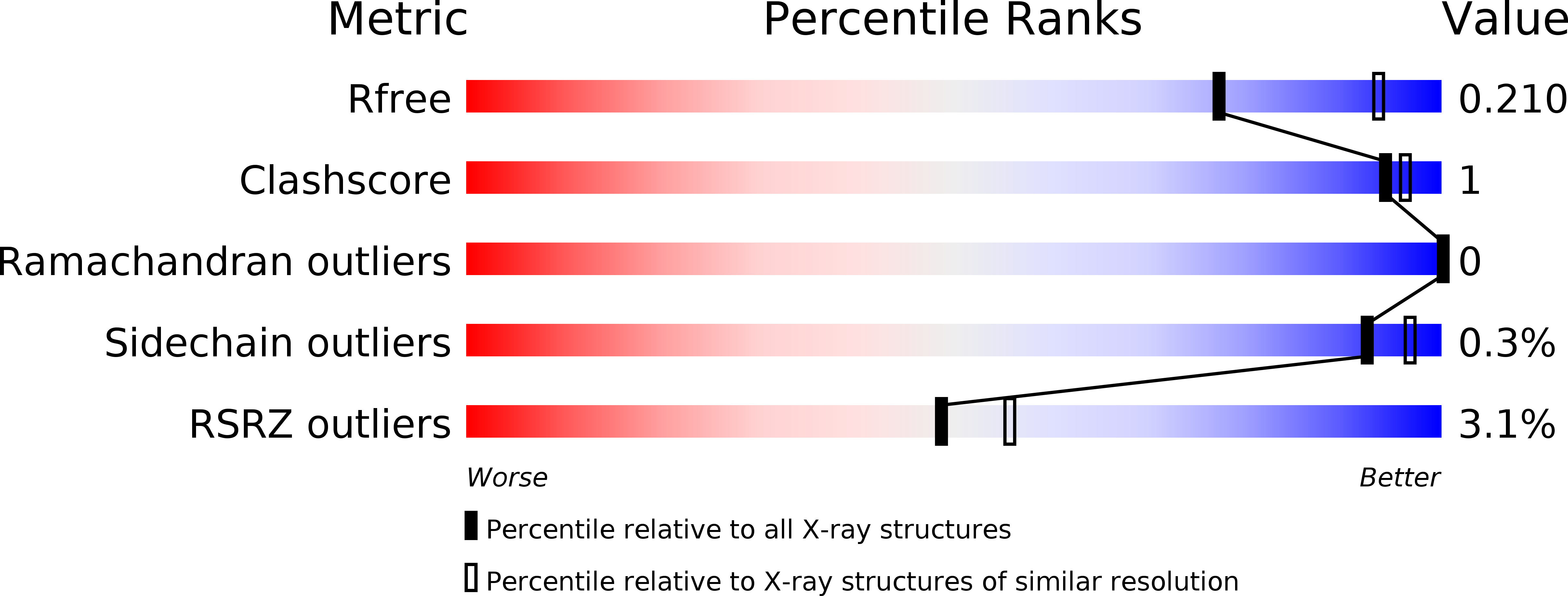
wwPDB Validation   3D Report Full Report


Ligand Structure Quality Assessment 


This is version 1.3 of the entry. See complete history


Literature

Structural characterization of an aldo-keto reductase (AKR2E5) from the silkworm Bombyx mori

Yamamoto, K.Higashiura, A.Suzuki, M.Shiotsuki, T.Sugahara, R.Fujii, T.Nakagawa, A.

(2016) Biochem Biophys Res Commun 474: 104-110

  • DOI: https://doi.org/10.1016/j.bbrc.2016.04.079
  • Primary Citation of Related Structures:  
    5AZ0, 5AZ1

  • PubMed Abstract: 

    We report a new member of the aldo-keto reductase (AKR) superfamily in the silkworm Bombyx mori. Based on its amino acid sequence, the new enzyme belongs to the AKR2 family and was previously assigned the systematic name AKR2E5. In the present study, recombinant AKR2E5 was expressed, purified to homogeneity, and characterized. The X-ray crystal structures were determined at 2.2 Å for the apoenzyme and at 2.3 Å resolution for the NADPH-AKR2E5 complex. Our results demonstrate that AKR2E5 is a 40-kDa monomer and includes the TIM- or (β/α)8-barrel typical for other AKRs. We found that AKR2E5 uses NADPH as a cosubstrate to reduce carbonyl compounds such as DL-glyceraldehyde, xylose, 3-hydroxy benzaldehyde, 17α-hydroxy progesterone, 11-hexadecenal, and bombykal. No NADH-dependent activity was detected. Site-directed mutagenesis of AKR2E5 indicates that amino acid residues Asp70, Tyr75, Lys104, and His137 contribute to catalytic activity, which is consistent with the data on other AKRs. To the best of our knowledge, AKR2E5 is only the second AKR characterized in silkworm. Our data should contribute to further understanding of the functional activity of insect AKRs.


  • Organizational Affiliation

    Kyushu University Graduate School, 6-10-1 Hakozaki, Higashi-ku, Fukuoka 812-8581, Japan. Electronic address: yamamok@agr.kyushu-u.ac.jp.


Macromolecules
Find similar proteins by:  (by identity cutoff)  |  3D Structure
Entity ID: 1
MoleculeChains Sequence LengthOrganismDetailsImage
Uncharacterized protein353Bombyx moriMutation(s): 0 
Gene Names: LOC101740031
Entity Groups  
Sequence Clusters30% Identity50% Identity70% Identity90% Identity95% Identity100% Identity
Sequence Annotations
Expand
  • Reference Sequence
Small Molecules
Ligands 4 Unique
IDChains Name / Formula / InChI Key2D Diagram3D Interactions
NDP
Query on NDP

Download Ideal Coordinates CCD File 
J [auth A]NADPH DIHYDRO-NICOTINAMIDE-ADENINE-DINUCLEOTIDE PHOSPHATE
C21 H30 N7 O17 P3
ACFIXJIJDZMPPO-NNYOXOHSSA-N
EDO
Query on EDO

Download Ideal Coordinates CCD File 
C [auth A]1,2-ETHANEDIOL
C2 H6 O2
LYCAIKOWRPUZTN-UHFFFAOYSA-N
ACT
Query on ACT

Download Ideal Coordinates CCD File 
D [auth A]
E [auth A]
F [auth A]
G [auth A]
H [auth A]
D [auth A],
E [auth A],
F [auth A],
G [auth A],
H [auth A],
I [auth A]
ACETATE ION
C2 H3 O2
QTBSBXVTEAMEQO-UHFFFAOYSA-M
CA
Query on CA

Download Ideal Coordinates CCD File 
B [auth A]CALCIUM ION
Ca
BHPQYMZQTOCNFJ-UHFFFAOYSA-N
Experimental Data & Validation

Experimental Data

  • Method: X-RAY DIFFRACTION
  • Resolution: 2.30 Å
  • R-Value Free: 0.206 
  • R-Value Work: 0.159 
  • R-Value Observed: 0.161 
  • Space Group: P 61 2 2
Unit Cell:
Length ( Å )Angle ( ˚ )
a = 111.952α = 90
b = 111.952β = 90
c = 147.651γ = 120
Software Package:
Software NamePurpose
REFMACrefinement
HKL-2000data reduction
HKL-2000data scaling
HKL-2000phasing

Structure Validation

View Full Validation Report


Ligand Structure Quality Assessment 


Entry History 

Deposition Data

Revision History  (Full details and data files)

  • Version 1.0: 2016-02-10
    Type: Initial release
  • Version 1.1: 2016-05-18
    Changes: Database references
  • Version 1.2: 2020-02-26
    Changes: Data collection, Database references, Derived calculations
  • Version 1.3: 2024-03-20
    Changes: Data collection, Database references