5AWQ | pdb_00005awq

Arthrobacter globiformis T6 isomalto-dextranse complexed with panose


Experimental Data Snapshot

  • Method: X-RAY DIFFRACTION
  • Resolution: 1.48 Å
  • R-Value Free: 
    0.187 (Depositor), 0.189 (DCC) 
  • R-Value Work: 
    0.161 (Depositor), 0.163 (DCC) 
  • R-Value Observed: 
    0.163 (Depositor) 

wwPDB Validation 3D Report Full Report

Validation slider image for 5AWQ

This is version 2.1 of the entry. See complete history

Literature

Crystal Structure and Mutational Analysis of Isomalto-dextranase, a Member of Glycoside Hydrolase Family 27

Okazawa, Y.Miyazaki, T.Yokoi, G.Ishizaki, Y.Nishikawa, A.Tonozuka, T.

(2015) J Biological Chem 290: 26339-26349

  • DOI: https://doi.org/10.1074/jbc.M115.680942
  • Primary Citation Related Structures: 
    5AWO, 5AWP, 5AWQ

  • PubMed Abstract: 

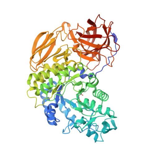
    Arthrobacter globiformis T6 isomalto-dextranase (AgIMD) is an enzyme that liberates isomaltose from the non-reducing end of a polymer of glucose, dextran. AgIMD is classified as a member of the glycoside hydrolase family (GH) 27, which comprises mainly α-galactosidases and α-N-acetylgalactosaminidases, whereas AgIMD does not show α-galactosidase or α-N-acetylgalactosaminidase activities. Here, we determined the crystal structure of AgIMD. AgIMD consists of the following three domains: A, C, and D. Domains A and C are identified as a (β/α)8-barrel catalytic domain and an antiparallel β-structure, respectively, both of which are commonly found in GH27 enzymes. However, domain A of AgIMD has subdomain B, loop-1, and loop-2, all of which are not found in GH27 human α-galactosidase. AgIMD in a complex with trisaccharide panose shows that Asp-207, a residue in loop-1, is involved in subsite +1. Kinetic parameters of the wild-type and mutant enzymes for the small synthetic saccharide p-nitrophenyl α-isomaltoside and the polysaccharide dextran were compared, showing that Asp-207 is important for the catalysis of dextran. Domain D is classified as carbohydrate-binding module (CBM) 35, and an isomaltose molecule is seen in this domain in the AgIMD-isomaltose complex. Domain D is highly homologous to CBM35 domains found in GH31 and GH66 enzymes. The results here indicate that some features found in GH13, -31, and -66 enzymes, such as subdomain B, residues at the subsite +1, and the CBM35 domain, are also observed in the GH27 enzyme AgIMD and thus provide insights into the evolutionary relationships among GH13, -27, -31, -36, and -66 enzymes.


  • Organizational Affiliation
    • From the Department of Applied Biological Science, Tokyo University of Agriculture and Technology, Tokyo 183-8509, Japan.

Macromolecule Content 

  • Total Structure Weight: 67.83 kDa 
  • Atom Count: 5,504 
  • Modeled Residue Count: 596 
  • Deposited Residue Count: 610 
  • Unique protein chains: 1

Macromolecules

Find similar proteins by:|  3D Structure
Entity ID: 1
MoleculeChains  Sequence LengthOrganismDetailsImage
Isomaltodextranase610Arthrobacter globiformisMutation(s): 0 
Gene Names: g2d
UniProt
Find proteins for Q7WSN5 (Arthrobacter globiformis)
Explore Q7WSN5 
Go to UniProtKB:  Q7WSN5
Entity Groups
Sequence Clusters30% Identity50% Identity70% Identity90% Identity95% Identity100% Identity
UniProt GroupQ7WSN5
Sequence Annotations
Expand
Reference Sequence

Oligosaccharides

Help  
Entity ID: 2
MoleculeChains Length2D Diagram GlycosylationD Interactions
alpha-D-glucopyranose-(1-6)-alpha-D-glucopyranose-(1-4)-alpha-D-glucopyranose
B
3N/A
Glycosylation Resources
GlyTouCan: G05704WW
GlyCosmos: G05704WW
GlyGen: G05704WW
Entity ID: 3
MoleculeChains Length2D Diagram GlycosylationD Interactions
alpha-D-glucopyranose-(1-6)-alpha-D-glucopyranose
C
2N/A
Glycosylation Resources
GlyTouCan: G69864PN
GlyCosmos: G69864PN

Small Molecules

Ligands 1 Unique
IDChains Name / Formula / InChI Key2D Diagram3D Interactions
PO4

Query on PO4


Download:Ideal Coordinates CCD File
D [auth A]PHOSPHATE ION
O4 P
NBIIXXVUZAFLBC-UHFFFAOYSA-K
Modified Residues  1 Unique
IDChains TypeFormula2D DiagramParent
SME
Query on SME
A
L-PEPTIDE LINKINGC5 H11 N O3 SMET

Experimental Data & Validation

Experimental Data

  • Method: X-RAY DIFFRACTION
  • Resolution: 1.48 Å
  • R-Value Free:  0.187 (Depositor), 0.189 (DCC) 
  • R-Value Work:  0.161 (Depositor), 0.163 (DCC) 
  • R-Value Observed: 0.163 (Depositor) 
Space Group: I 41
Unit Cell:
Length ( Å )Angle ( ˚ )
a = 123.878α = 90
b = 123.878β = 90
c = 85.261γ = 90
Software Package:
Software NamePurpose
REFMACrefinement
HKL-2000data reduction
HKL-2000data scaling
MOLREPphasing

Structure Validation

View Full Validation Report


Entry History 

Deposition Data

  • Released Date: 2015-09-09 
  • Deposition Author(s): Tonozuka, T.

Revision History  (Full details and data files)

  • Version 1.0: 2015-09-09
    Type: Initial release
  • Version 1.1: 2015-10-21
    Changes: Database references
  • Version 1.2: 2015-11-04
    Changes: Structure summary
  • Version 1.3: 2015-11-11
    Changes: Database references
  • Version 1.4: 2020-02-26
    Changes: Data collection, Database references, Derived calculations
  • Version 2.0: 2020-07-29
    Type: Remediation
    Reason: Carbohydrate remediation
    Changes: Atomic model, Data collection, Derived calculations, Structure summary
  • Version 2.1: 2025-04-09
    Changes: Data collection, Database references, Derived calculations, Structure summary