5AI3 | pdb_00005ai3

X-ray structure of 113Cd-substituted Perdeuterated Pyrococcus furiosus rubredoxin to 1.02A resolution at 295K in a quartz capillary


Experimental Data Snapshot

  • Method: X-RAY DIFFRACTION
  • Resolution: 1.02 Å
  • R-Value Free: 
    0.126 (Depositor), 0.136 (DCC) 
  • R-Value Work: 
    0.106 (Depositor), 0.110 (DCC) 
  • R-Value Observed: 
    0.107 (Depositor) 

wwPDB Validation 3D Report Full Report

Validation slider image for 5AI3

This is version 1.3 of the entry. See complete history

Literature

Macromolecular Structure Phasing by Neutron Anomalous Diffraction.

Cuypers, M.G.Mason, S.A.Mossou, E.Haertlein, M.Forsyth, V.T.Mitchell, E.P.

(2016) Sci Rep 6: 31487

  • DOI: https://doi.org/10.1038/srep31487
  • Primary Citation Related Structures: 
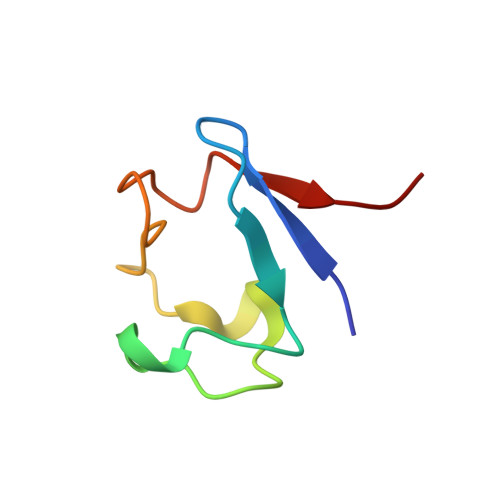
    5AI2, 5AI3

  • PubMed Abstract: 

    In this report we show for the first time that neutron anomalous dispersion can be used in a practical manner to determine experimental phases of a protein crystal structure, providing a new tool for structural biologists. The approach is demonstrated through the use of a state-of-the-art monochromatic neutron diffractometer at the Institut Laue-Langevin (ILL) in combination with crystals of perdeuterated protein that minimise the level of hydrogen incoherent scattering and enhance the visibility of the anomalous signal. The protein used was rubredoxin in which cadmium replaced the iron at the iron-sulphur site. While this study was carried out using a steady-state neutron beam source, the results will be of major interest for capabilities at existing and emerging spallation neutron sources where time-of-flight instruments provide inherent energy discrimination. In particular this capability may be expected to offer unique opportunities to a rapidly developing structural biology community where there is increasing interest in the identification of protonation states, protein/water interactions and protein-ligand interactions - all of which are of central importance to a wide range of fundamental and applied areas in the biosciences.


  • Organizational Affiliation
    • Faculty of Natural Sciences, Keele University, Staffordshire, ST5 5BG, United Kingdom.

Macromolecule Content 

  • Total Structure Weight: 6.14 kDa 
  • Atom Count: 569 
  • Modeled Residue Count: 54 
  • Deposited Residue Count: 54 
  • Unique protein chains: 1

Macromolecules

Find similar proteins by:|  3D Structure
Entity ID: 1
MoleculeChains  Sequence LengthOrganismDetailsImage
RUBREDOXIN54Pyrococcus furiosusMutation(s): 0 
UniProt
Find proteins for P24297 (Pyrococcus furiosus (strain ATCC 43587 / DSM 3638 / JCM 8422 / Vc1))
Explore P24297 
Go to UniProtKB:  P24297
Entity Groups
Sequence Clusters30% Identity50% Identity70% Identity90% Identity95% Identity100% Identity
UniProt GroupP24297
Sequence Annotations
Expand
Reference Sequence

Small Molecules

Ligands 1 Unique
IDChains Name / Formula / InChI Key2D Diagram3D Interactions
CD

Query on CD


Download:Ideal Coordinates CCD File
B [auth A]CADMIUM ION
Cd
WLZRMCYVCSSEQC-UHFFFAOYSA-N

Experimental Data & Validation

Experimental Data

  • Method: X-RAY DIFFRACTION
  • Resolution: 1.02 Å
  • R-Value Free:  0.126 (Depositor), 0.136 (DCC) 
  • R-Value Work:  0.106 (Depositor), 0.110 (DCC) 
  • R-Value Observed: 0.107 (Depositor) 
Space Group: P 21 21 21
Unit Cell:
Length ( Å )Angle ( ˚ )
a = 34.443α = 90
b = 35.141β = 90
c = 43.785γ = 90
Software Package:
Software NamePurpose
PHENIXrefinement
XDSdata reduction
SCALAdata scaling
MOLREPphasing

Structure Validation

View Full Validation Report


Entry History 

Deposition Data

Revision History  (Full details and data files)

  • Version 1.0: 2016-03-02
    Type: Initial release
  • Version 1.1: 2016-03-16
    Changes: Atomic model
  • Version 1.2: 2016-08-24
    Changes: Database references
  • Version 1.3: 2024-05-08
    Changes: Data collection, Database references, Derived calculations, Other