5AFB | pdb_00005afb

Crystal structure of the Latrophilin3 Lectin and Olfactomedin Domains


Experimental Data Snapshot

  • Method: X-RAY DIFFRACTION
  • Resolution: 2.16 Å
  • R-Value Free: 
    0.245 (Depositor), 0.248 (DCC) 
  • R-Value Work: 
    0.204 (Depositor), 0.212 (DCC) 
  • R-Value Observed: 
    0.206 (Depositor) 

wwPDB Validation 3D Report Full Report

Validation slider image for 5AFB

Ligand Structure Quality Assessment 


This is version 1.4 of the entry. See complete history

Literature

Structural Basis of Latrophilin-Flrt Interaction.

Jackson, V.A.Del Toro, D.Carrasquero, M.Roversi, P.Harlos, K.Klein, R.Seiradake, E.

(2015) Structure 23: 774

  • DOI: https://doi.org/10.1016/j.str.2015.01.013
  • Primary Citation Related Structures: 
    5AFB

  • PubMed Abstract: 

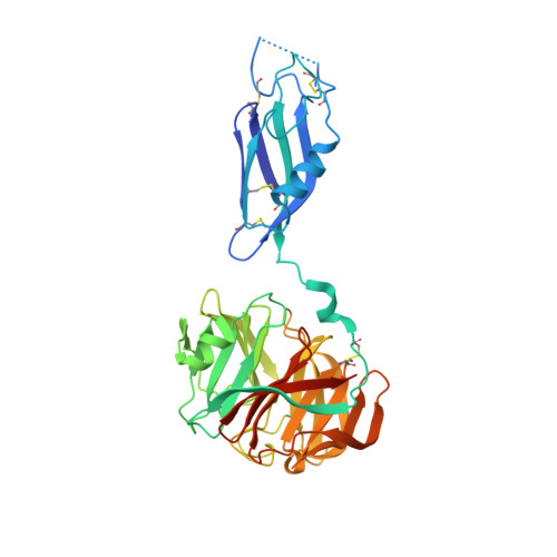
    Latrophilins, receptors for spider venom α-latrotoxin, are adhesion type G-protein-coupled receptors with emerging functions in synapse development. The N-terminal region binds the endogenous cell adhesion molecule FLRT, a major regulator of cortical and synapse development. We present crystallographic data for the mouse Latrophilin3 lectin and olfactomedin-like (Olf) domains, thereby revealing the Olf β-propeller fold and conserved calcium-binding site. We locate the FLRT-Latrophilin binding surfaces by a combination of sequence conservation analysis, point mutagenesis, and surface plasmon resonance experiments. In stripe assays, we show that wild-type Latrophilin3 and its high-affinity interactor FLRT2, but not the binding-impaired mutants we generated, promote HeLa cell adhesion. In contrast, cortical neurons expressing endogenous FLRTs are repelled by wild-type Latrophilin3 and not by the binding-impaired mutant. Taken together, we present molecular level insights into Latrophilin structure, its FLRT-binding mechanism, and a role for Latrophilin and FLRT that goes beyond a simply adhesive interaction.


  • Organizational Affiliation
    • Department of Biochemistry, Oxford University, South Parks Road, Oxford OX1 3QU, UK.

Macromolecule Content 

  • Total Structure Weight: 44.51 kDa 
  • Atom Count: 2,930 
  • Modeled Residue Count: 347 
  • Deposited Residue Count: 384 
  • Unique protein chains: 1

Macromolecules

Find similar proteins by:|  3D Structure
Entity ID: 1
MoleculeChains  Sequence LengthOrganismDetailsImage
LATROPHILIN-3384Homo sapiensMutation(s): 0 
UniProt & NIH Common Fund Data Resources
Find proteins for Q9HAR2 (Homo sapiens)
Explore Q9HAR2 
Go to UniProtKB:  Q9HAR2
PHAROS:  Q9HAR2
GTEx:  ENSG00000150471 
Entity Groups
Sequence Clusters30% Identity50% Identity70% Identity90% Identity95% Identity100% Identity
UniProt GroupQ9HAR2
Glycosylation
Glycosylation Sites: 1Go to GlyGen: Q9HAR2-1
Sequence Annotations
Expand
Reference Sequence

Experimental Data & Validation

Experimental Data

  • Method: X-RAY DIFFRACTION
  • Resolution: 2.16 Å
  • R-Value Free:  0.245 (Depositor), 0.248 (DCC) 
  • R-Value Work:  0.204 (Depositor), 0.212 (DCC) 
  • R-Value Observed: 0.206 (Depositor) 
Space Group: I 2 2 2
Unit Cell:
Length ( Å )Angle ( ˚ )
a = 78.43α = 90
b = 96.65β = 90
c = 101.64γ = 90
Software Package:
Software NamePurpose
BUSTERrefinement
xia2data reduction
xia2data scaling
autoSHARPphasing

Structure Validation

View Full Validation Report


Ligand Structure Quality Assessment 


Entry History 

Deposition Data

Revision History  (Full details and data files)

  • Version 1.0: 2015-03-18
    Type: Initial release
  • Version 1.1: 2015-04-22
    Changes: Database references
  • Version 1.2: 2016-03-09
    Changes: Database references, Other, Structure summary
  • Version 1.3: 2020-07-29
    Type: Remediation
    Reason: Carbohydrate remediation
    Changes: Data collection, Derived calculations, Other, Structure summary
  • Version 1.4: 2024-10-09
    Changes: Data collection, Database references, Structure summary