5ADV | pdb_00005adv

The Periplasmic Binding Protein CeuE of Campylobacter jejuni preferentially binds the iron(III) complex of the Linear Dimer Component of Enterobactin


Experimental Data Snapshot

  • Method: X-RAY DIFFRACTION
  • Resolution: 2.10 Å
  • R-Value Free: 
    0.260 (Depositor), 0.260 (DCC) 
  • R-Value Work: 
    0.218 (Depositor), 0.223 (DCC) 
  • R-Value Observed: 
    0.220 (Depositor) 

Starting Model: experimental
View more details

wwPDB Validation 3D Report Full Report

Validation slider image for 5ADV

Ligand Structure Quality Assessment 


This is version 1.2 of the entry. See complete history

Literature

Bacteria in an Intense Competition for Iron: Key Component of the Campylobacter Jejuni Iron Uptake System Scavenges Enterobactin Hydrolysis Product.

Raines, D.J.Moroz, O.V.Blagova, E.V.Turkenburg, J.P.Wilson, K.S.Duhme-Klair, A.

(2016) Proc Natl Acad Sci U S A 113: 5850

  • DOI: https://doi.org/10.1073/pnas.1520829113
  • Primary Citation Related Structures: 
    5ADV, 5ADW

  • PubMed Abstract: 

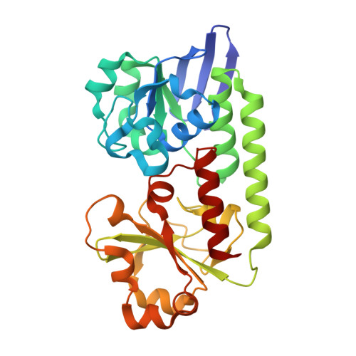
    To acquire essential Fe(III), bacteria produce and secrete siderophores with high affinity and selectivity for Fe(III) to mediate its uptake into the cell. Here, we show that the periplasmic binding protein CeuE of Campylobacter jejuni, which was previously thought to bind the Fe(III) complex of the hexadentate siderophore enterobactin (Kd ∼ 0.4 ± 0.1 µM), preferentially binds the Fe(III) complex of the tetradentate enterobactin hydrolysis product bis(2,3-dihydroxybenzoyl-l-Ser) (H5-bisDHBS) (Kd = 10.1 ± 3.8 nM). The protein selects Λ-configured [Fe(bisDHBS)](2-) from a pool of diastereomeric Fe(III)-bisDHBS species that includes complexes with metal-to-ligand ratios of 1:1 and 2:3. Cocrystal structures show that, in addition to electrostatic interactions and hydrogen bonding, [Fe(bisDHBS)](2-) binds through coordination of His227 and Tyr288 to the iron center. Similar binding is observed for the Fe(III) complex of the bidentate hydrolysis product 2,3-dihydroxybenzoyl-l-Ser, [Fe(monoDHBS)2](3-) The mutation of His227 and Tyr288 to noncoordinating residues (H227L/Y288F) resulted in a substantial loss of affinity for [Fe(bisDHBS)](2-) (Kd ∼ 0.5 ± 0.2 µM). These results suggest a previously unidentified role for CeuE within the Fe(III) uptake system of C. jejuni, provide a molecular-level understanding of the underlying binding pocket adaptations, and rationalize reports on the use of enterobactin hydrolysis products by C. jejuni, Vibrio cholerae, and other bacteria with homologous periplasmic binding proteins.


  • Organizational Affiliation
    • Department of Chemistry, University of York, Heslington, York YO10 5DD, United Kingdom;

Macromolecule Content 

  • Total Structure Weight: 97.01 kDa 
  • Atom Count: 6,851 
  • Modeled Residue Count: 861 
  • Deposited Residue Count: 861 
  • Unique protein chains: 1

Macromolecules

Find similar proteins by:|  3D Structure
Entity ID: 1
MoleculeChains  Sequence LengthOrganismDetailsImage
ENTEROCHELIN UPTAKE PERIPLASMIC BINDING PROTEIN
A, B, C
287Campylobacter jejuniMutation(s): 0 
UniProt
Find proteins for Q0P8Q4 (Campylobacter jejuni subsp. jejuni serotype O:2 (strain ATCC 700819 / NCTC 11168))
Explore Q0P8Q4 
Go to UniProtKB:  Q0P8Q4
Entity Groups
Sequence Clusters30% Identity50% Identity70% Identity90% Identity95% Identity100% Identity
UniProt GroupQ0P8Q4
Sequence Annotations
Expand
Reference Sequence

Small Molecules

Ligands 4 Unique
IDChains Name / Formula / InChI Key2D Diagram3D Interactions
EHS

Query on EHS


Download:Ideal Coordinates CCD File
K [auth B]2S-2-[(2,3-DIHYDROXYPHENYL)CARBONYLAMINO]-3-[(2S)-2-[(2,3-DIHYDROXYPHENYL)CARBONYLAMINO]-3-HYDROXY-PROPANOYL]OXY-PROPANOIC ACID
C20 H20 N2 O11
KLXJDVFEFZPIMN-RYUDHWBXSA-N
DBS

Query on DBS


Download:Ideal Coordinates CCD File
E [auth A],
F [auth A],
M [auth C],
N [auth C]
2-(2,3-DIHYDROXY-BENZOYLAMINO)-3-HYDROXY-PROPIONIC ACID
C10 H11 N O6
VDTYHTVHFIIEIL-LURJTMIESA-N
DMS

Query on DMS


Download:Ideal Coordinates CCD File
G [auth A],
H [auth B],
I [auth B]
DIMETHYL SULFOXIDE
C2 H6 O S
IAZDPXIOMUYVGZ-UHFFFAOYSA-N
FE

Query on FE


Download:Ideal Coordinates CCD File
D [auth A],
J [auth B],
L [auth C]
FE (III) ION
Fe
VTLYFUHAOXGGBS-UHFFFAOYSA-N

Experimental Data & Validation

Experimental Data

  • Method: X-RAY DIFFRACTION
  • Resolution: 2.10 Å
  • R-Value Free:  0.260 (Depositor), 0.260 (DCC) 
  • R-Value Work:  0.218 (Depositor), 0.223 (DCC) 
  • R-Value Observed: 0.220 (Depositor) 
Space Group: P 1
Unit Cell:
Length ( Å )Angle ( ˚ )
a = 58.07α = 83.09
b = 63.09β = 76.9
c = 67.16γ = 79.21
Software Package:
Software NamePurpose
REFMACrefinement
xia2data reduction

Structure Validation

View Full Validation Report


Ligand Structure Quality Assessment 


Entry History 

Deposition Data

Revision History  (Full details and data files)

  • Version 1.0: 2016-05-25
    Type: Initial release
  • Version 1.1: 2016-06-08
    Changes: Database references
  • Version 1.2: 2024-01-10
    Changes: Data collection, Database references, Derived calculations, Other, Refinement description