5AB2 | pdb_00005ab2

Crystal structure of aminopeptidase ERAP2 with ligand


Experimental Data Snapshot

  • Method: X-RAY DIFFRACTION
  • Resolution: 2.73 Å
  • R-Value Free: 
    0.286 (Depositor) 
  • R-Value Work: 
    0.223 (Depositor) 
  • R-Value Observed: 
    0.226 (Depositor) 

Starting Model: experimental
View more details

wwPDB Validation 3D Report Full Report

Validation slider image for 5AB2

Ligand Structure Quality Assessment 


This is version 2.2 of the entry. See complete history

Literature

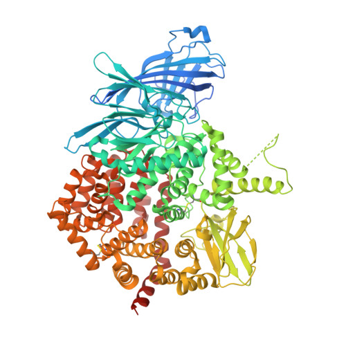
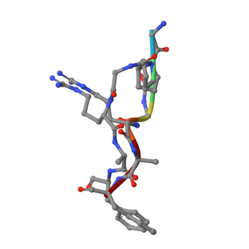
Structural Basis for Antigenic Peptide Recognition and Processing by Endoplasmic Reticulum (Er) Aminopeptidase 2.

Mpakali, A.Giastas, P.Mathioudakis, N.Mavridis, I.M.Saridakis, E.Stratikos, E.

(2015) J Biological Chem 290: 26021

  • DOI: https://doi.org/10.1074/jbc.M115.685909
  • Primary Citation Related Structures: 
    5AB0, 5AB2, 5CU5

  • PubMed Abstract: 

    Endoplasmic reticulum (ER) aminopeptidases process antigenic peptide precursors to generate epitopes for presentation by MHC class I molecules and help shape the antigenic peptide repertoire and cytotoxic T-cell responses. To perform this function, ER aminopeptidases have to recognize and process a vast variety of peptide sequences. To understand how these enzymes recognize substrates, we determined crystal structures of ER aminopeptidase 2 (ERAP2) in complex with a substrate analogue and a peptidic product to 2.5 and 2.7 Å, respectively, and compared them to the apo-form structure determined to 3.0 Å. The peptides were found within the internal cavity of the enzyme with no direct access to the outside solvent. The substrate analogue extends away from the catalytic center toward the distal end of the internal cavity, making interactions with several shallow pockets along the path. A similar configuration was evident for the peptidic product, although decreasing electron density toward its C terminus indicated progressive disorder. Enzymatic analysis confirmed that visualized interactions can either positively or negatively impact in vitro trimming rates. Opportunistic side-chain interactions and lack of deep specificity pockets support a limited-selectivity model for antigenic peptide processing by ERAP2. In contrast to proposed models for the homologous ERAP1, no specific recognition of the peptide C terminus by ERAP2 was evident, consistent with functional differences in length selection and self-activation between these two enzymes. Our results suggest that ERAP2 selects substrates by sequestering them in its internal cavity and allowing opportunistic interactions to determine trimming rates, thus combining substrate permissiveness with sequence bias.


  • Organizational Affiliation
    • From the National Center for Scientific Research Demokritos, Agia Paraskevi, Athens 15310, Greece.

Macromolecule Content 

  • Total Structure Weight: 230.87 kDa 
  • Atom Count: 15,144 
  • Modeled Residue Count: 1,788 
  • Deposited Residue Count: 1,946 
  • Unique protein chains: 2

Macromolecules

Find similar proteins by:|  3D Structure
Entity ID: 1
MoleculeChains  Sequence LengthOrganismDetailsImage
ENDOPLASMIC RETICULUM AMINOPEPTIDASE 2
A, B
967Homo sapiensMutation(s): 0 
EC: 3.4.11
UniProt & NIH Common Fund Data Resources
Find proteins for Q6P179 (Homo sapiens)
Explore Q6P179 
Go to UniProtKB:  Q6P179
PHAROS:  Q6P179
GTEx:  ENSG00000164308 
Entity Groups
Sequence Clusters30% Identity50% Identity70% Identity90% Identity95% Identity100% Identity
UniProt GroupQ6P179
Glycosylation
Glycosylation Sites: 6Go to GlyGen: Q6P179-1
Sequence Annotations
Expand
Reference Sequence
Find similar proteins by:  Sequence   |   3D Structure  
Entity ID: 2
MoleculeChains  Sequence LengthOrganismDetailsImage
GPI
C, D
6synthetic constructMutation(s): 0 

Oligosaccharides

Help  
Entity ID: 3
MoleculeChains Length2D Diagram GlycosylationD Interactions
beta-D-mannopyranose-(1-4)-2-acetamido-2-deoxy-beta-D-glucopyranose-(1-4)-2-acetamido-2-deoxy-beta-D-glucopyranose
E, G
3N-Glycosylation
Glycosylation Resources
GlyTouCan: G15407YE
GlyCosmos: G15407YE
GlyGen: G15407YE
Entity ID: 4
MoleculeChains Length2D Diagram GlycosylationD Interactions
2-acetamido-2-deoxy-beta-D-glucopyranose-(1-4)-2-acetamido-2-deoxy-beta-D-glucopyranose
F, H, I, J, K
F, H, I, J, K, M, N
2N-Glycosylation
Glycosylation Resources
GlyTouCan: G42666HT
GlyCosmos: G42666HT
GlyGen: G42666HT
Entity ID: 5
MoleculeChains Length2D Diagram GlycosylationD Interactions
beta-D-mannopyranose-(1-6)-beta-D-mannopyranose-(1-4)-2-acetamido-2-deoxy-beta-D-glucopyranose-(1-4)-2-acetamido-2-deoxy-beta-D-glucopyranose
L
4N-Glycosylation
Glycosylation Resources
GlyTouCan: G40894EI
GlyCosmos: G40894EI
GlyGen: G40894EI

Small Molecules

Ligands 3 Unique
IDChains Name / Formula / InChI Key2D Diagram3D Interactions
NAG

Query on NAG


Download:Ideal Coordinates CCD File
O [auth A],
P [auth A],
U [auth B],
V [auth B],
W [auth B]
2-acetamido-2-deoxy-beta-D-glucopyranose
C8 H15 N O6
OVRNDRQMDRJTHS-FMDGEEDCSA-N
ZN

Query on ZN


Download:Ideal Coordinates CCD File
Q [auth A],
X [auth B]
ZINC ION
Zn
PTFCDOFLOPIGGS-UHFFFAOYSA-N
EDO

Query on EDO


Download:Ideal Coordinates CCD File
AA [auth B]
R [auth A]
S [auth A]
T [auth A]
Y [auth B]
AA [auth B],
R [auth A],
S [auth A],
T [auth A],
Y [auth B],
Z [auth B]
1,2-ETHANEDIOL
C2 H6 O2
LYCAIKOWRPUZTN-UHFFFAOYSA-N

Experimental Data & Validation

Experimental Data

  • Method: X-RAY DIFFRACTION
  • Resolution: 2.73 Å
  • R-Value Free:  0.286 (Depositor) 
  • R-Value Work:  0.223 (Depositor) 
  • R-Value Observed: 0.226 (Depositor) 
Space Group: P 1 21 1
Unit Cell:
Length ( Å )Angle ( ˚ )
a = 75.65α = 90
b = 135.465β = 90.24
c = 128.212γ = 90
Software Package:
Software NamePurpose
PHENIXrefinement
MOSFLMdata reduction
SCALAdata scaling
MOLREPphasing

Structure Validation

View Full Validation Report


Ligand Structure Quality Assessment 


Entry History 

Deposition Data

Revision History  (Full details and data files)

  • Version 1.0: 2015-09-30
    Type: Initial release
  • Version 1.1: 2015-11-11
    Changes: Database references
  • Version 1.2: 2019-04-03
    Changes: Data collection, Derived calculations, Other, Source and taxonomy
  • Version 2.0: 2020-07-29
    Type: Remediation
    Reason: Carbohydrate remediation
    Changes: Atomic model, Data collection, Derived calculations, Other, Structure summary
  • Version 2.1: 2024-01-10
    Changes: Data collection, Database references, Derived calculations, Refinement description, Structure summary
  • Version 2.2: 2024-10-09
    Changes: Structure summary