5A5U | pdb_00005a5u

Structure of mammalian eIF3 in the context of the 43S preinitiation complex


Experimental Data Snapshot

  • Method: ELECTRON MICROSCOPY
  • Resolution: 9.00 Å
  • Aggregation State: PARTICLE 
  • Reconstruction Method: SINGLE PARTICLE 

wwPDB Validation 3D Report Full Report

Validation slider image for 5A5U

This is version 1.5 of the entry. See complete history

Literature

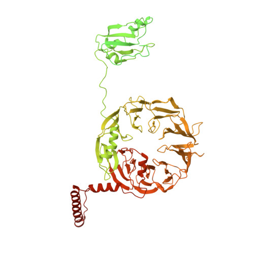
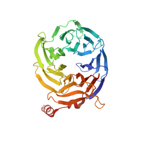
Structure of Mammalian Eif3 in the Context of the 43S Preinitiation Complex.

Des-Georges, A.Dhote, V.Kuhn, L.Hellen, C.U.T.Pestova, T.V.Frank, J.Hashem, Y.

(2015) Nature 525: 491

  • DOI: https://doi.org/10.1038/nature14891
  • Primary Citation Related Structures: 
    5A5T, 5A5U

  • PubMed Abstract: 

    During eukaryotic translation initiation, 43S complexes, comprising a 40S ribosomal subunit, initiator transfer RNA and initiation factors (eIF) 2, 3, 1 and 1A, attach to the 5'-terminal region of messenger RNA and scan along it to the initiation codon. Scanning on structured mRNAs also requires the DExH-box protein DHX29. Mammalian eIF3 contains 13 subunits and participates in nearly all steps of translation initiation. Eight subunits having PCI (proteasome, COP9 signalosome, eIF3) or MPN (Mpr1, Pad1, amino-terminal) domains constitute the structural core of eIF3, to which five peripheral subunits are flexibly linked. Here we present a cryo-electron microscopy structure of eIF3 in the context of the DHX29-bound 43S complex, showing the PCI/MPN core at ∼6 Å resolution. It reveals the organization of the individual subunits and their interactions with components of the 43S complex. We were able to build near-complete polyalanine-level models of the eIF3 PCI/MPN core and of two peripheral subunits. The implications for understanding mRNA ribosomal attachment and scanning are discussed.


  • Organizational Affiliation
    • HHMI, Department of Biochemistry and Molecular Biophysics, Columbia University, New York, New York 10032, USA.

Macromolecule Content 

  • Total Structure Weight: 167.82 kDa 
  • Atom Count: 8,124 
  • Modeled Residue Count: 1,009 
  • Deposited Residue Count: 1,522 
  • Unique protein chains: 3

Macromolecules

Find similar proteins by:|  3D Structure
Entity ID: 1
MoleculeChains  Sequence LengthOrganismDetailsImage
EUKARYOTIC INITIATION FACTOR 354Oryctolagus cuniculusMutation(s): 0 
Find similar proteins by:|  3D Structure
Entity ID: 2
MoleculeChains  Sequence LengthOrganismDetailsImage
EUKARYOTIC TRANSLATION INITIATION FACTOR 3 SUBUNIT B1,121Oryctolagus cuniculusMutation(s): 0 
UniProt
Find proteins for G1SZ03 (Oryctolagus cuniculus)
Explore G1SZ03 
Go to UniProtKB:  G1SZ03
Entity Groups
Sequence Clusters30% Identity50% Identity70% Identity90% Identity95% Identity100% Identity
UniProt GroupG1SZ03
Sequence Annotations
Expand
Reference Sequence
Find similar proteins by:|  3D Structure
Entity ID: 3
MoleculeChains  Sequence LengthOrganismDetailsImage
EUKARYOTIC TRANSLATION INITIATION FACTOR 3 SUBUNIT IC [auth I]347Oryctolagus cuniculusMutation(s): 0 
UniProt
Find proteins for P40217 (Saccharomyces cerevisiae (strain ATCC 204508 / S288c))
Explore P40217 
Go to UniProtKB:  P40217
Entity Groups
Sequence Clusters30% Identity50% Identity70% Identity90% Identity95% Identity100% Identity
UniProt GroupP40217
Sequence Annotations
Expand
Reference Sequence

Experimental Data & Validation

Experimental Data

  • Method: ELECTRON MICROSCOPY
  • Resolution: 9.00 Å
  • Aggregation State: PARTICLE 
  • Reconstruction Method: SINGLE PARTICLE 
EM Software:
TaskSoftware PackageVersion
RECONSTRUCTIONRELION

Structure Validation

View Full Validation Report


Entry History 

Deposition Data

Revision History  (Full details and data files)

  • Version 1.0: 2015-09-09
    Type: Initial release
  • Version 1.1: 2015-09-23
    Changes: Database references
  • Version 1.2: 2015-09-30
    Changes: Database references
  • Version 1.3: 2017-08-30
    Changes: Data collection
  • Version 1.4: 2018-10-03
    Changes: Data collection
  • Version 1.5: 2024-05-08
    Changes: Data collection, Database references