


Crystal Structure of Human Spindlin3
Srikannathasan, V., Gileadi, C., Johansson, C., Shrestha, L., Tallon, R., Burgess-Brown, N.A., von Delft, F., Arrowsmith, C.H., Bountra, C., Edwards, A., Oppermann, U.To be published.
Experimental Data Snapshot
Starting Model: experimental
View more details
wwPDB Validation 3D Report Full Report

Entity ID: 1 | |||||
|---|---|---|---|---|---|
| Molecule | Chains | Sequence Length | Organism | Details | Image |
| SPINDLIN-3 | 222 | Homo sapiens | Mutation(s): 0 | 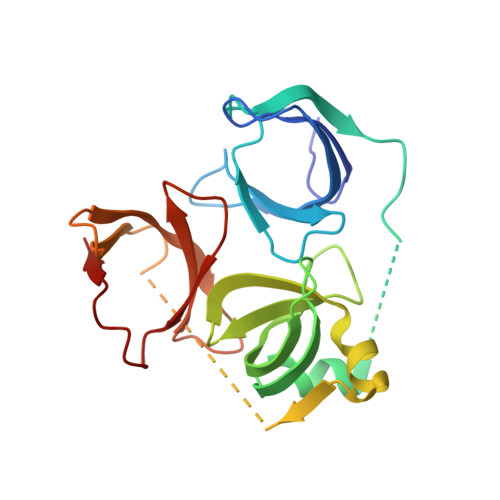 | |
UniProt & NIH Common Fund Data Resources | |||||
PHAROS: Q5JUX0 GTEx: ENSG00000204271 | |||||
Entity Groups | |||||
| Sequence Clusters | 30% Identity50% Identity70% Identity90% Identity95% Identity100% Identity | ||||
| UniProt Group | Q5JUX0 | ||||
Sequence AnnotationsExpand | |||||
Reference Sequence | |||||
| Length ( Å ) | Angle ( ˚ ) |
|---|---|
| a = 58.58 | α = 90 |
| b = 129.4 | β = 90 |
| c = 129.48 | γ = 90 |
| Software Name | Purpose |
|---|---|
| REFMAC | refinement |
| MOSFLM | data reduction |
| SCALA | data scaling |
| CCP4I | phasing |


