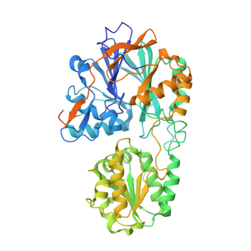
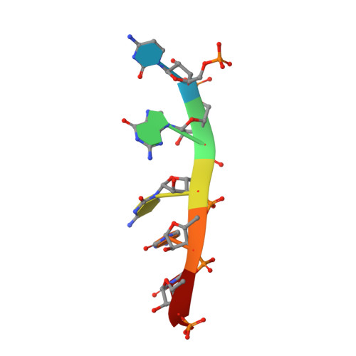
5A0T

Catalysis and 5' end sensing by ribonuclease RNase J of the metallo- beta-lactamase family


Experimental Data Snapshot

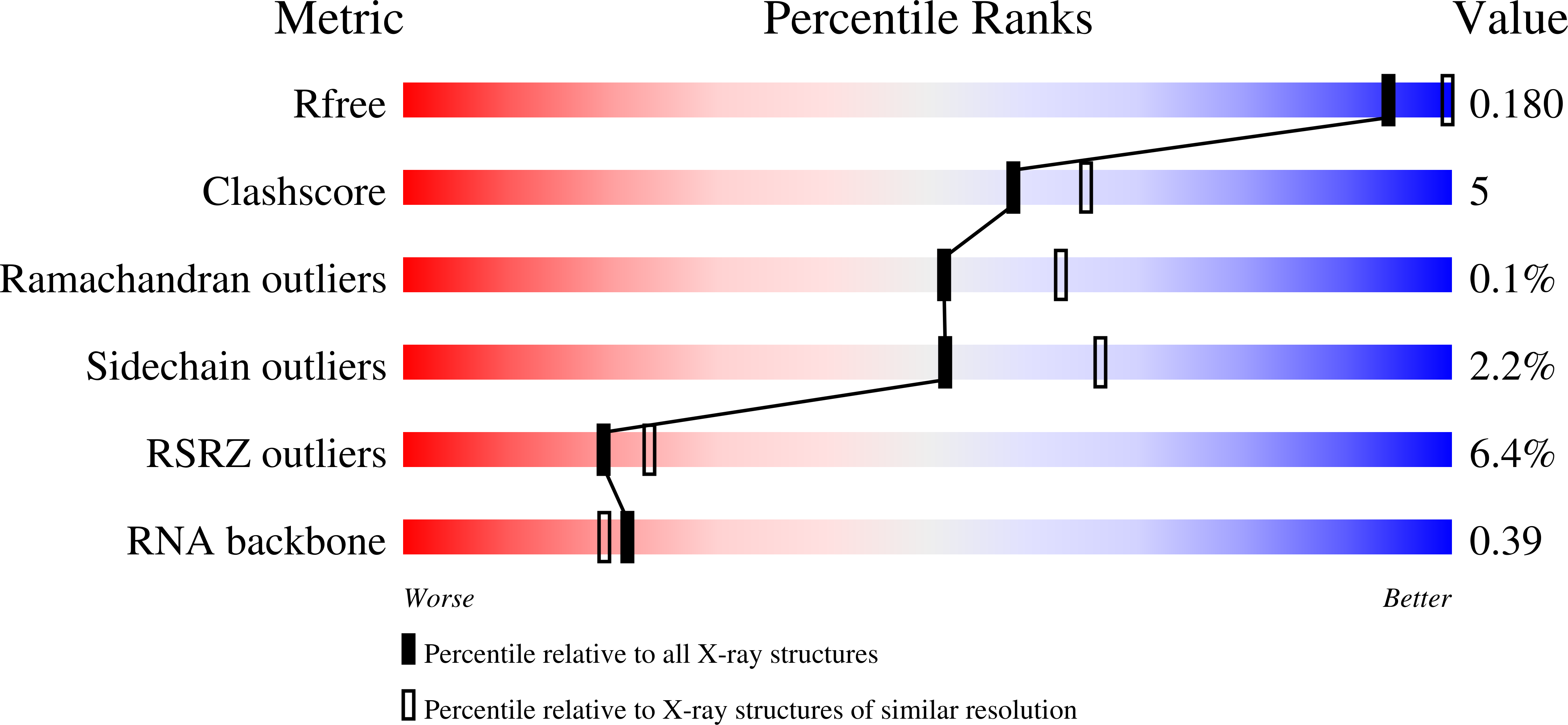
  • Method: X-RAY DIFFRACTION
  • Resolution: 2.28 Å
  • R-Value Free: 0.177 
  • R-Value Work: 0.145 
  • R-Value Observed: 0.146 

Starting Model: experimental
View more details

wwPDB Validation   3D Report Full Report


This is version 1.2 of the entry. See complete history


Literature

Linkage of Catalysis and 5' End Recognition in Ribonuclease Rnase J

Pei, X.Y.Bralley, P.Jones, G.H.Luisi, B.F.

(2015) Nucleic Acids Res 43: 8066

  • DOI: https://doi.org/10.1093/nar/gkv732
  • Primary Citation of Related Structures:  
    5A0T, 5A0V

  • PubMed Abstract: 

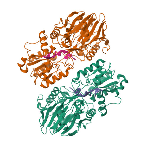
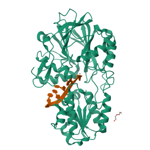
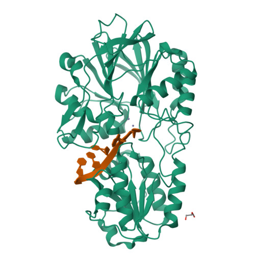
    In diverse bacterial species, the turnover and processing of many RNAs is mediated by the ribonuclease RNase J, a member of the widely occurring metallo-β-lactamase enzyme family. We present crystal structures of Streptomyces coelicolor RNase J with bound RNA in pre- and post-cleavage states, at 2.27 Å and 2.80 Å resolution, respectively. These structures reveal snapshots of the enzyme cleaving substrate directionally and sequentially from the 5' terminus. In the pre-cleavage state, a water molecule is coordinated to a zinc ion pair in the active site but is imperfectly oriented to launch a nucleophilic attack on the phosphate backbone. A conformational switch is envisaged that enables the in-line positioning of the attacking water and may be facilitated by magnesium ions. Adjacent to the scissile bond, four bases are stacked in a tightly sandwiching pocket, and mutagenesis results indicate that this organization helps to drive processive exo-ribonucleolytic cleavage. Like its numerous homologues, S. coelicolor RNase J can also cleave some RNA internally, and the structural data suggest how the preference for exo- versus endo-cleavage mode is linked with recognition of the chemical status of the substrate's 5' end.


  • Organizational Affiliation

    Department of Biochemistry, University of Cambridge, Tennis Court Road, Cambridge CB2 1GA, UK.


Macromolecules

Find similar proteins by:  (by identity cutoff)  |  3D Structure
Entity ID: 1
MoleculeChains Sequence LengthOrganismDetailsImage
RIBONUCLEASE J
A, B
561Streptomyces coelicolor A3(2)Mutation(s): 0 
EC: 3.1
UniProt
Find proteins for O86842 (Streptomyces coelicolor (strain ATCC BAA-471 / A3(2) / M145))
Explore O86842 
Go to UniProtKB:  O86842
Entity Groups  
Sequence Clusters30% Identity50% Identity70% Identity90% Identity95% Identity100% Identity
UniProt GroupO86842
Sequence Annotations
Expand
  • Reference Sequence

Find similar nucleic acids by:  Sequence   |   3D Structure  

Entity ID: 2
MoleculeChains LengthOrganismImage
5'-R(*CP*GP*CP*CP*UP)-3'C [auth E],
D [auth F]
6Escherichia coli BL21(DE3)
Sequence Annotations
Expand
  • Reference Sequence
Experimental Data & Validation

Experimental Data

  • Method: X-RAY DIFFRACTION
  • Resolution: 2.28 Å
  • R-Value Free: 0.177 
  • R-Value Work: 0.145 
  • R-Value Observed: 0.146 
  • Space Group: P 43 2 2
Unit Cell:
Length ( Å )Angle ( ˚ )
a = 186.187α = 90
b = 186.187β = 90
c = 113.382γ = 90
Software Package:
Software NamePurpose
MOSFLMdata reduction
Aimlessdata scaling
SHELXphasing
PHENIXphasing
PHENIXrefinement

Structure Validation

View Full Validation Report


Entry History 

Deposition Data

Revision History  (Full details and data files)

  • Version 1.0: 2015-08-19
    Type: Initial release
  • Version 1.1: 2015-10-07
    Changes: Database references
  • Version 1.2: 2024-01-10
    Changes: Data collection, Database references, Derived calculations, Other, Refinement description