4ZN3 | pdb_00004zn3

Crystal Structure of MjSpt4:Spt5 complex conformation B


Experimental Data Snapshot

  • Method: X-RAY DIFFRACTION
  • Resolution: 2.30 Å
  • R-Value Free: 
    0.256 (Depositor), 0.261 (DCC) 
  • R-Value Work: 
    0.227 (Depositor), 0.227 (DCC) 
  • R-Value Observed: 
    0.229 (Depositor) 

Starting Model: experimental
View more details

wwPDB Validation 3D Report Full Report

Validation slider image for 4ZN3

This is version 1.1 of the entry. See complete history

Literature

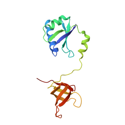
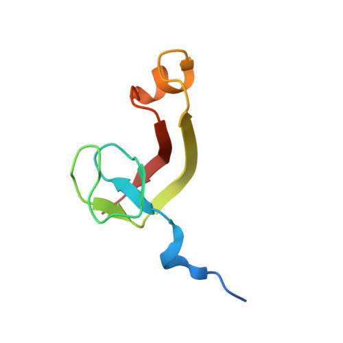
Structural and biochemical insights into the DNA-binding mode of MjSpt4p:Spt5 complex at the exit tunnel of RNAPII

Guo, G.R.Gao, Y.X.Zhu, Z.L.Zhao, D.Liu, Z.Zhou, H.H.Niu, L.W.Teng, M.K.

(2015) J Struct Biol 192: 418-425

  • DOI: https://doi.org/10.1016/j.jsb.2015.09.023
  • Primary Citation Related Structures: 
    4ZN1, 4ZN3

  • PubMed Abstract: 

    Spt5 (NusG in bacteria) is the only RNA polymerase-associated factor known to be conserved in all three domains of life. In archaea and eukaryotes, Spt5 associates with Spt4, an elongation factor that is absent in bacteria, to form a functional heterodimeric complex. Previous studies suggest that the Spt4:Spt5 complex interacts directly with DNA at the double-stranded DNA exit tunnel of RNA polymerase to regulate gene transcription. In this study, the DNA-binding ability of Spt4:Spt5 from the archaeon Methanocaldococcus jannaschii was confirmed via nuclear magnetic resonance chemical shift perturbation and fluorescence polarization assays. Crystallographic analysis of the full-length MjSpt4:Spt5 revealed two distinct conformations of the C-terminal KOW domain of Spt5. A similar alkaline region was found on the Spt4:Spt5 surface in both crystal forms, and identified as double-stranded DNA binding patch through mutagenesis-fluorescence polarization assays. Based on these structural and biochemical data, the Spt4:Spt5-DNA binding model was built for the first time.


  • Organizational Affiliation
    • Hefei National Laboratory for Physical Sciences at the Microscale and School of Life Sciences, University of Science and Technology of China, Hefei, Anhui 230026, People's Republic of China; Key Laboratory of Structural Biology, Chinese Academy of Sciences, Hefei, Anhui 230026, People's Republic of China.

Macromolecule Content 

  • Total Structure Weight: 24.6 kDa 
  • Atom Count: 1,693 
  • Modeled Residue Count: 214 
  • Deposited Residue Count: 219 
  • Unique protein chains: 2

Macromolecules

Find similar proteins by:|  3D Structure
Entity ID: 1
MoleculeChains  Sequence LengthOrganismDetailsImage
Transcription elongation factor Spt5147Methanocaldococcus jannaschii DSM 2661Mutation(s): 0 
Gene Names: spt5
UniProt
Find proteins for Q57818 (Methanocaldococcus jannaschii (strain ATCC 43067 / DSM 2661 / JAL-1 / JCM 10045 / NBRC 100440))
Explore Q57818 
Go to UniProtKB:  Q57818
Entity Groups
Sequence Clusters30% Identity50% Identity70% Identity90% Identity95% Identity100% Identity
UniProt GroupQ57818
Sequence Annotations
Expand
Reference Sequence
Find similar proteins by:|  3D Structure
Entity ID: 2
MoleculeChains  Sequence LengthOrganismDetailsImage
Transcription elongation factor Spt472Methanocaldococcus jannaschii DSM 2661Mutation(s): 0 
Gene Names: spt4
UniProt
Find proteins for Q57839 (Methanocaldococcus jannaschii (strain ATCC 43067 / DSM 2661 / JAL-1 / JCM 10045 / NBRC 100440))
Explore Q57839 
Go to UniProtKB:  Q57839
Entity Groups
Sequence Clusters30% Identity50% Identity70% Identity90% Identity95% Identity100% Identity
UniProt GroupQ57839
Sequence Annotations
Expand
Reference Sequence

Experimental Data & Validation

Experimental Data

  • Method: X-RAY DIFFRACTION
  • Resolution: 2.30 Å
  • R-Value Free:  0.256 (Depositor), 0.261 (DCC) 
  • R-Value Work:  0.227 (Depositor), 0.227 (DCC) 
  • R-Value Observed: 0.229 (Depositor) 
Space Group: P 21 21 21
Unit Cell:
Length ( Å )Angle ( ˚ )
a = 45.395α = 90
b = 88.943β = 90
c = 91.955γ = 90
Software Package:
Software NamePurpose
REFMACrefinement
HKL-2000data scaling
PHASERphasing

Structure Validation

View Full Validation Report


Entry History 

Deposition Data

Revision History  (Full details and data files)

  • Version 1.0: 2016-03-16
    Type: Initial release
  • Version 1.1: 2023-11-08
    Changes: Data collection, Database references, Derived calculations, Refinement description