4ZN3

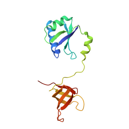
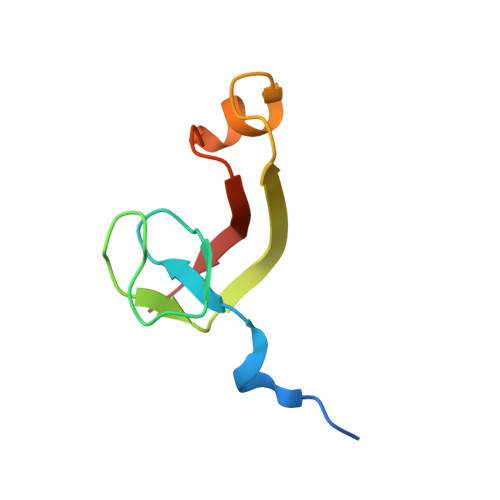
Crystal Structure of MjSpt4:Spt5 complex conformation B


Experimental Data Snapshot

  • Method: X-RAY DIFFRACTION
  • Resolution: 2.30 Å
  • R-Value Free: 0.256 
  • R-Value Work: 0.227 
  • R-Value Observed: 0.229 

Starting Model: experimental
View more details

wwPDB Validation   3D Report Full Report


This is version 1.1 of the entry. See complete history


Literature

Structural and biochemical insights into the DNA-binding mode of MjSpt4p:Spt5 complex at the exit tunnel of RNAPII

Guo, G.R.Gao, Y.X.Zhu, Z.L.Zhao, D.Liu, Z.Zhou, H.H.Niu, L.W.Teng, M.K.

(2015) J Struct Biol 192: 418-425

  • DOI: https://doi.org/10.1016/j.jsb.2015.09.023
  • Primary Citation of Related Structures:  
    4ZN1, 4ZN3

  • PubMed Abstract: 

    Spt5 (NusG in bacteria) is the only RNA polymerase-associated factor known to be conserved in all three domains of life. In archaea and eukaryotes, Spt5 associates with Spt4, an elongation factor that is absent in bacteria, to form a functional heterodimeric complex. Previous studies suggest that the Spt4:Spt5 complex interacts directly with DNA at the double-stranded DNA exit tunnel of RNA polymerase to regulate gene transcription. In this study, the DNA-binding ability of Spt4:Spt5 from the archaeon Methanocaldococcus jannaschii was confirmed via nuclear magnetic resonance chemical shift perturbation and fluorescence polarization assays. Crystallographic analysis of the full-length MjSpt4:Spt5 revealed two distinct conformations of the C-terminal KOW domain of Spt5. A similar alkaline region was found on the Spt4:Spt5 surface in both crystal forms, and identified as double-stranded DNA binding patch through mutagenesis-fluorescence polarization assays. Based on these structural and biochemical data, the Spt4:Spt5-DNA binding model was built for the first time.


  • Organizational Affiliation

    Hefei National Laboratory for Physical Sciences at the Microscale and School of Life Sciences, University of Science and Technology of China, Hefei, Anhui 230026, People's Republic of China; Key Laboratory of Structural Biology, Chinese Academy of Sciences, Hefei, Anhui 230026, People's Republic of China.


Macromolecules
Find similar proteins by:  (by identity cutoff)  |  3D Structure
Entity ID: 1
MoleculeChains Sequence LengthOrganismDetailsImage
Transcription elongation factor Spt5147Methanocaldococcus jannaschii DSM 2661Mutation(s): 0 
Gene Names: spt5
UniProt
Find proteins for Q57818 (Methanocaldococcus jannaschii (strain ATCC 43067 / DSM 2661 / JAL-1 / JCM 10045 / NBRC 100440))
Explore Q57818 
Go to UniProtKB:  Q57818
Entity Groups  
Sequence Clusters30% Identity50% Identity70% Identity90% Identity95% Identity100% Identity
UniProt GroupQ57818
Sequence Annotations
Expand
  • Reference Sequence
Find similar proteins by:  (by identity cutoff)  |  3D Structure
Entity ID: 2
MoleculeChains Sequence LengthOrganismDetailsImage
Transcription elongation factor Spt472Methanocaldococcus jannaschii DSM 2661Mutation(s): 0 
Gene Names: spt4
UniProt
Find proteins for Q57839 (Methanocaldococcus jannaschii (strain ATCC 43067 / DSM 2661 / JAL-1 / JCM 10045 / NBRC 100440))
Explore Q57839 
Go to UniProtKB:  Q57839
Entity Groups  
Sequence Clusters30% Identity50% Identity70% Identity90% Identity95% Identity100% Identity
UniProt GroupQ57839
Sequence Annotations
Expand
  • Reference Sequence
Experimental Data & Validation

Experimental Data

  • Method: X-RAY DIFFRACTION
  • Resolution: 2.30 Å
  • R-Value Free: 0.256 
  • R-Value Work: 0.227 
  • R-Value Observed: 0.229 
  • Space Group: P 21 21 21
Unit Cell:
Length ( Å )Angle ( ˚ )
a = 45.395α = 90
b = 88.943β = 90
c = 91.955γ = 90
Software Package:
Software NamePurpose
REFMACrefinement
HKL-2000data scaling
PHASERphasing

Structure Validation

View Full Validation Report


Entry History 

Deposition Data

Revision History  (Full details and data files)

  • Version 1.0: 2016-03-16
    Type: Initial release
  • Version 1.1: 2023-11-08
    Changes: Data collection, Database references, Derived calculations, Refinement description