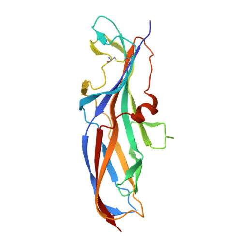
4Z3E

Crystal structure of the lectin domain of PapG from E. coli BI47 in complex with SSEA4 in space group P212121


Experimental Data Snapshot

  • Method: X-RAY DIFFRACTION
  • Resolution: 1.80 Å
  • R-Value Free: 0.224 
  • R-Value Work: 0.178 
  • R-Value Observed: 0.180 

wwPDB Validation   3D Report Full Report


This is version 2.1 of the entry. See complete history


Literature

Carbohydrate-Lectin Interactions: An Unexpected Contribution to Affinity.

Navarra, G.Zihlmann, P.Jakob, R.P.Stangier, K.Preston, R.C.Rabbani, S.Smiesko, M.Wagner, B.Maier, T.Ernst, B.

(2017) Chembiochem 18: 539-544

  • DOI: https://doi.org/10.1002/cbic.201600615
  • Primary Citation of Related Structures:  
    4Z3E

  • PubMed Abstract: 

    Uropathogenic E. coli exploit PapG-II adhesin for infecting host cells of the kidney; the expression of PapG-II at the tip of bacterial pili correlates with the onset of pyelonephritis in humans, a potentially life-threatening condition. It was envisaged that blocking PapG-II (and thus bacterial adhesion) would provide a viable therapeutic alternative to conventional antibiotic treatment. In our search for potent PapG-II antagonists, we observed an increase in affinity when tetrasaccharide 1, the natural ligand of PapG-II in human kidneys, was elongated to hexasaccharide 2, even though the additional Siaα(2-3)Gal extension is not in direct contact with the lectin. ITC studies suggest that the increased affinity results from partial desolvation of nonbinding regions of the hexasaccharide; this is ultimately responsible for perturbation of the outer hydration layers. Our results are in agreement with previous observations and suggest a general mechanism for modulating carbohydrate-protein interactions based on nonbinding regions of the ligand.


  • Organizational Affiliation

    Institute of Molecular Pharmacy, Pharmacenter, University of Basel, Klingelbergstrasse 50, 4056, Basel, Switzerland.


Macromolecules
Find similar proteins by:  (by identity cutoff)  |  3D Structure
Entity ID: 1
MoleculeChains Sequence LengthOrganismDetailsImage
PapG, lectin domain198Escherichia coliMutation(s): 0 
UniProt
Find proteins for A0A182DW20 (Escherichia coli)
Explore A0A182DW20 
Go to UniProtKB:  A0A182DW20
Entity Groups  
Sequence Clusters30% Identity50% Identity70% Identity90% Identity95% Identity100% Identity
UniProt GroupA0A182DW20
Sequence Annotations
Expand
  • Reference Sequence
Oligosaccharides

Help

Entity ID: 2
MoleculeChains Length2D Diagram Glycosylation3D Interactions
beta-D-galactopyranose-(1-3)-2-acetamido-2-deoxy-beta-D-galactopyranose-(1-3)-alpha-D-galactopyranose-(1-4)-beta-D-galactopyranose-(1-4)-beta-D-glucopyranose
B
5N/A
Glycosylation Resources
GlyTouCan:  G99053DY
GlyCosmos:  G99053DY
GlyGen:  G99053DY
Experimental Data & Validation

Experimental Data

  • Method: X-RAY DIFFRACTION
  • Resolution: 1.80 Å
  • R-Value Free: 0.224 
  • R-Value Work: 0.178 
  • R-Value Observed: 0.180 
  • Space Group: P 21 21 21
Unit Cell:
Length ( Å )Angle ( ˚ )
a = 48.21α = 90
b = 52.22β = 90
c = 76.32γ = 90
Software Package:
Software NamePurpose
REFMACrefinement
XDSdata reduction
XSCALEdata scaling
PHASERphasing

Structure Validation

View Full Validation Report


Entry History 

Deposition Data

Revision History  (Full details and data files)

  • Version 1.0: 2016-04-13
    Type: Initial release
  • Version 1.1: 2017-01-25
    Changes: Database references
  • Version 1.2: 2017-02-15
    Changes: Database references
  • Version 1.3: 2017-03-29
    Changes: Database references
  • Version 2.0: 2020-07-29
    Type: Remediation
    Reason: Carbohydrate remediation
    Changes: Atomic model, Data collection, Derived calculations, Structure summary
  • Version 2.1: 2024-01-10
    Changes: Data collection, Database references, Derived calculations, Refinement description, Structure summary