4XPJ | pdb_00004xpj

Crystal structure of Nerve growth factor in complex with lysophosphatidylinositol


Experimental Data Snapshot

  • Method: X-RAY DIFFRACTION
  • Resolution: 2.60 Å
  • R-Value Free: 
    0.266 (Depositor), 0.271 (DCC) 
  • R-Value Work: 
    0.195 (Depositor), 0.200 (DCC) 
  • R-Value Observed: 
    0.199 (Depositor) 

Starting Model: experimental
View more details

wwPDB Validation 3D Report Full Report

Validation slider image for 4XPJ

Ligand Structure Quality Assessment 


This is version 1.2 of the entry. See complete history

Literature

The structure of nerve growth factor in complex with lysophosphatidylinositol

Sun, H.L.Jiang, T.

(2015) Acta Crystallogr F Struct Biol Commun 71: 906-912

  • DOI: https://doi.org/10.1107/S2053230X15008870
  • Primary Citation Related Structures: 
    4XPJ

  • PubMed Abstract: 

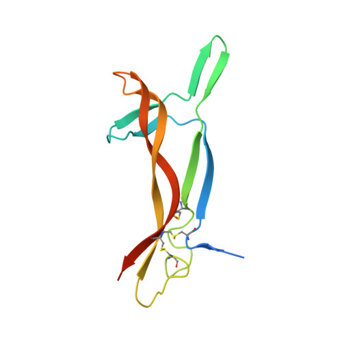
    Nerve growth factor (NGF) is an important protein that is involved in a variety of physiological processes in cell survival, differentiation, proliferation and maintenance. The previously reported crystal structure of mouse NGF (mNGF) in complex with lysophosphatidylserine (LysoPS) showed that mNGF can bind LysoPS at its dimeric interface. To expand the understanding of the structural basis for specific lipid recognition by NGF, the crystal structure of mNGF complexed with lysophosphatidylinositol (13:0 LysoPI) was solved. Interestingly, in addition to Lys88, which interacts with the head glycerol group and the phosphate group of LysoPI, as seen in the mNGF-LysoPS structure, two additional residues, Tyr52 and Arg50, were found to assist in lipid binding by forming hydrogen bonds to the inositol moiety of the LysoPI molecule. The results suggest a specific recognition mechanism of inositol group-containing lipids by NGF, which may help in the design of bioactive compounds that can be delivered by NGF.


  • Organizational Affiliation
    • National Laboratory of Biomacromolecules, Institute of Biophysics, Chinese Academy of Sciences, 15 Datun Road, Beijing 100101, People's Republic of China.

Macromolecule Content 

  • Total Structure Weight: 27.51 kDa 
  • Atom Count: 1,750 
  • Modeled Residue Count: 210 
  • Deposited Residue Count: 240 
  • Unique protein chains: 1

Macromolecules

Find similar proteins by:|  3D Structure
Entity ID: 1
MoleculeChains  Sequence LengthOrganismDetailsImage
Beta-nerve growth factor
A, B
120Mus musculusMutation(s): 0 
UniProt
Find proteins for P01139 (Mus musculus)
Explore P01139 
Go to UniProtKB:  P01139
Entity Groups
Sequence Clusters30% Identity50% Identity70% Identity90% Identity95% Identity100% Identity
UniProt GroupP01139
Sequence Annotations
Expand
Reference Sequence

Small Molecules

Ligands 1 Unique
IDChains Name / Formula / InChI Key2D Diagram3D Interactions
LPY

Query on LPY


Download:Ideal Coordinates CCD File
C [auth B](2R)-2-hydroxy-3-{[(S)-hydroxy{[(1S,2R,3R,4S,5S,6R)-2,3,4,5,6-pentahydroxycyclohexyl]oxy}phosphoryl]oxy}propyl tridecanoate
C22 H43 O12 P
QGSQXOJTKFYHRK-FBYKMOGESA-N

Experimental Data & Validation

Experimental Data

  • Method: X-RAY DIFFRACTION
  • Resolution: 2.60 Å
  • R-Value Free:  0.266 (Depositor), 0.271 (DCC) 
  • R-Value Work:  0.195 (Depositor), 0.200 (DCC) 
  • R-Value Observed: 0.199 (Depositor) 
Space Group: H 3 2
Unit Cell:
Length ( Å )Angle ( ˚ )
a = 96.689α = 90
b = 96.689β = 90
c = 164.339γ = 120
Software Package:
Software NamePurpose
PHENIXrefinement
HKL-2000data processing
HKL-2000data scaling
AMoREphasing

Structure Validation

View Full Validation Report


Ligand Structure Quality Assessment 


Entry History 

Deposition Data

Revision History  (Full details and data files)

  • Version 1.0: 2015-07-15
    Type: Initial release
  • Version 1.1: 2023-11-08
    Changes: Data collection, Database references, Derived calculations, Refinement description, Source and taxonomy, Structure summary
  • Version 1.2: 2024-11-13
    Changes: Structure summary