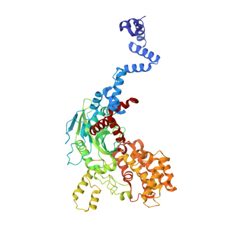
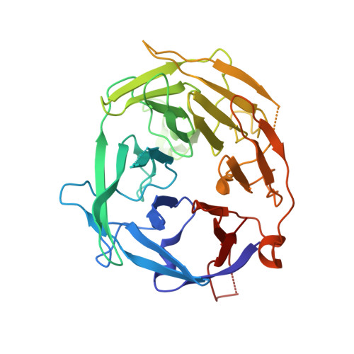
4WWX

Crystal structure of the core RAG1/2 recombinase


Experimental Data Snapshot

  • Method: X-RAY DIFFRACTION
  • Resolution: 3.20 Å
  • R-Value Free: 0.259 
  • R-Value Work: 0.205 
  • R-Value Observed: 0.208 

wwPDB Validation   3D Report Full Report


This is version 1.5 of the entry. See complete history


Literature

Crystal structure of the V(D)J recombinase RAG1-RAG2.

Kim, M.S.Lapkouski, M.Yang, W.Gellert, M.

(2015) Nature 518: 507-511

  • DOI: https://doi.org/10.1038/nature14174
  • Primary Citation of Related Structures:  
    4WWX

  • PubMed Abstract: 

    V(D)J recombination in the vertebrate immune system generates a highly diverse population of immunoglobulins and T-cell receptors by combinatorial joining of segments of coding DNA. The RAG1-RAG2 protein complex initiates this site-specific recombination by cutting DNA at specific sites flanking the coding segments. Here we report the crystal structure of the mouse RAG1-RAG2 complex at 3.2 Å resolution. The 230-kilodalton RAG1-RAG2 heterotetramer is 'Y-shaped', with the amino-terminal domains of the two RAG1 chains forming an intertwined stalk. Each RAG1-RAG2 heterodimer composes one arm of the 'Y', with the active site in the middle and RAG2 at its tip. The RAG1-RAG2 structure rationalizes more than 60 mutations identified in immunodeficient patients, as well as a large body of genetic and biochemical data. The architectural similarity between RAG1 and the hairpin-forming transposases Hermes and Tn5 suggests the evolutionary conservation of these DNA rearrangements.


  • Organizational Affiliation

    Laboratory of Molecular Biology, NIDDK, NIH, Bethesda, Maryland 20892, USA.


Macromolecules
Find similar proteins by:  (by identity cutoff)  |  3D Structure
Entity ID: 1
MoleculeChains Sequence LengthOrganismDetailsImage
V(D)J recombination-activating protein 1A [auth B],
C [auth E]
618Mus musculusMutation(s): 0 
Gene Names: Rag1
EC: 3.1 (PDB Primary Data), 6.3.2 (PDB Primary Data), 2.3.2.27 (UniProt)
UniProt
Find proteins for P15919 (Mus musculus)
Explore P15919 
Go to UniProtKB:  P15919
Entity Groups  
Sequence Clusters30% Identity50% Identity70% Identity90% Identity95% Identity100% Identity
UniProt GroupP15919
Sequence Annotations
Expand
  • Reference Sequence
Find similar proteins by:  (by identity cutoff)  |  3D Structure
Entity ID: 2
MoleculeChains Sequence LengthOrganismDetailsImage
V(D)J recombination-activating protein 2B [auth X],
D [auth Y]
349Mus musculusMutation(s): 0 
Gene Names: Rag2Rag-2
UniProt
Find proteins for P21784 (Mus musculus)
Explore P21784 
Go to UniProtKB:  P21784
Entity Groups  
Sequence Clusters30% Identity50% Identity70% Identity90% Identity95% Identity100% Identity
UniProt GroupP21784
Sequence Annotations
Expand
  • Reference Sequence
Experimental Data & Validation

Experimental Data

  • Method: X-RAY DIFFRACTION
  • Resolution: 3.20 Å
  • R-Value Free: 0.259 
  • R-Value Work: 0.205 
  • R-Value Observed: 0.208 
  • Space Group: C 2 2 21
Unit Cell:
Length ( Å )Angle ( ˚ )
a = 168.76α = 90
b = 180.05β = 90
c = 200.19γ = 90
Software Package:
Software NamePurpose
PHENIXrefinement
PDB_EXTRACTdata extraction
XSCALEdata scaling
PHASERphasing

Structure Validation

View Full Validation Report


Entry History 

Deposition Data

Revision History  (Full details and data files)

  • Version 1.0: 2015-02-25
    Type: Initial release
  • Version 1.1: 2015-03-04
    Changes: Database references
  • Version 1.2: 2015-03-11
    Changes: Database references
  • Version 1.3: 2017-08-23
    Changes: Data collection, Database references, Derived calculations, Other, Source and taxonomy
  • Version 1.4: 2017-11-22
    Changes: Refinement description
  • Version 1.5: 2024-02-28
    Changes: Data collection, Database references