4WWX | pdb_00004wwx

Crystal structure of the core RAG1/2 recombinase


Experimental Data Snapshot

  • Method: X-RAY DIFFRACTION
  • Resolution: 3.20 Å
  • R-Value Free: 
    0.259 (Depositor), 0.268 (DCC) 
  • R-Value Work: 
    0.205 (Depositor), 0.213 (DCC) 
  • R-Value Observed: 
    0.208 (Depositor) 

wwPDB Validation 3D Report Full Report

Validation slider image for 4WWX

This is version 1.5 of the entry. See complete history

Literature

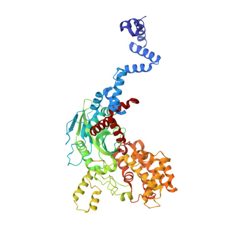
Crystal structure of the V(D)J recombinase RAG1-RAG2.

Kim, M.S.Lapkouski, M.Yang, W.Gellert, M.

(2015) Nature 518: 507-511

  • DOI: https://doi.org/10.1038/nature14174
  • Primary Citation Related Structures: 
    4WWX

  • PubMed Abstract: 

    V(D)J recombination in the vertebrate immune system generates a highly diverse population of immunoglobulins and T-cell receptors by combinatorial joining of segments of coding DNA. The RAG1-RAG2 protein complex initiates this site-specific recombination by cutting DNA at specific sites flanking the coding segments. Here we report the crystal structure of the mouse RAG1-RAG2 complex at 3.2 Å resolution. The 230-kilodalton RAG1-RAG2 heterotetramer is 'Y-shaped', with the amino-terminal domains of the two RAG1 chains forming an intertwined stalk. Each RAG1-RAG2 heterodimer composes one arm of the 'Y', with the active site in the middle and RAG2 at its tip. The RAG1-RAG2 structure rationalizes more than 60 mutations identified in immunodeficient patients, as well as a large body of genetic and biochemical data. The architectural similarity between RAG1 and the hairpin-forming transposases Hermes and Tn5 suggests the evolutionary conservation of these DNA rearrangements.


  • Organizational Affiliation
    • Laboratory of Molecular Biology, NIDDK, NIH, Bethesda, Maryland 20892, USA.

Macromolecule Content 

  • Total Structure Weight: 219.72 kDa 
  • Atom Count: 14,978 
  • Modeled Residue Count: 1,876 
  • Deposited Residue Count: 1,934 
  • Unique protein chains: 2

Macromolecules

Find similar proteins by:|  3D Structure
Entity ID: 1
MoleculeChains  Sequence LengthOrganismDetailsImage
V(D)J recombination-activating protein 1A [auth B],
C [auth E]
618Mus musculusMutation(s): 0 
Gene Names: Rag1
EC: 3.1 (PDB Primary Data), 6.3.2 (PDB Primary Data), 2.3.2.27 (UniProt)
UniProt
Find proteins for P15919 (Mus musculus)
Explore P15919 
Go to UniProtKB:  P15919
Entity Groups
Sequence Clusters30% Identity50% Identity70% Identity90% Identity95% Identity100% Identity
UniProt GroupP15919
Sequence Annotations
Expand
Reference Sequence
Find similar proteins by:|  3D Structure
Entity ID: 2
MoleculeChains  Sequence LengthOrganismDetailsImage
V(D)J recombination-activating protein 2B [auth X],
D [auth Y]
349Mus musculusMutation(s): 0 
Gene Names: Rag2Rag-2
UniProt
Find proteins for P21784 (Mus musculus)
Explore P21784 
Go to UniProtKB:  P21784
Entity Groups
Sequence Clusters30% Identity50% Identity70% Identity90% Identity95% Identity100% Identity
UniProt GroupP21784
Sequence Annotations
Expand
Reference Sequence

Experimental Data & Validation

Experimental Data

  • Method: X-RAY DIFFRACTION
  • Resolution: 3.20 Å
  • R-Value Free:  0.259 (Depositor), 0.268 (DCC) 
  • R-Value Work:  0.205 (Depositor), 0.213 (DCC) 
  • R-Value Observed: 0.208 (Depositor) 
Space Group: C 2 2 21
Unit Cell:
Length ( Å )Angle ( ˚ )
a = 168.76α = 90
b = 180.05β = 90
c = 200.19γ = 90
Software Package:
Software NamePurpose
PHENIXrefinement
PDB_EXTRACTdata extraction
XSCALEdata scaling
PHASERphasing

Structure Validation

View Full Validation Report


Entry History 

Deposition Data

Revision History  (Full details and data files)

  • Version 1.0: 2015-02-25
    Type: Initial release
  • Version 1.1: 2015-03-04
    Changes: Database references
  • Version 1.2: 2015-03-11
    Changes: Database references
  • Version 1.3: 2017-08-23
    Changes: Data collection, Database references, Derived calculations, Other, Source and taxonomy
  • Version 1.4: 2017-11-22
    Changes: Refinement description
  • Version 1.5: 2024-02-28
    Changes: Data collection, Database references