4WMK

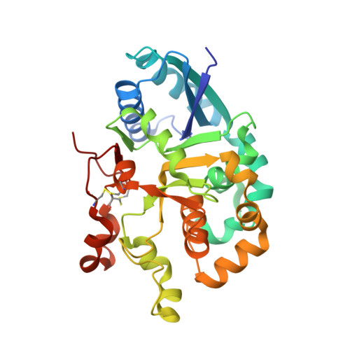
Crystal structure of mouse Xyloside xylosyltransferase 1 complexed with manganese, product ligand and UDP (Product complex II)


Experimental Data Snapshot

  • Method: X-RAY DIFFRACTION
  • Resolution: 2.08 Å
  • R-Value Free: 0.246 
  • R-Value Work: 0.191 
  • R-Value Observed: 0.194 

wwPDB Validation   3D Report Full Report


Ligand Structure Quality Assessment 


This is version 2.1 of the entry. See complete history


Literature

Notch-modifying xylosyltransferase structures support an SNi-like retaining mechanism.

Yu, H.Takeuchi, M.LeBarron, J.Kantharia, J.London, E.Bakker, H.Haltiwanger, R.S.Li, H.Takeuchi, H.

(2015) Nat Chem Biol 11: 847-854

  • DOI: https://doi.org/10.1038/nchembio.1927
  • Primary Citation of Related Structures:  
    4WLG, 4WLM, 4WLZ, 4WM0, 4WMA, 4WMB, 4WMI, 4WMK, 4WN2

  • PubMed Abstract: 

    A major question remaining in glycobiology is how a glycosyltransferase (GT) that retains the anomeric linkage of a sugar catalyzes the reaction. Xyloside α-1,3-xylosyltransferase (XXYLT1) is a retaining GT that regulates Notch receptor activation by adding xylose to the Notch extracellular domain. Here, using natural acceptor and donor substrates and active Mus musculus XXYLT1, we report a series of crystallographic snapshots along the reaction, including an unprecedented natural and competent Michaelis reaction complex for retaining enzymes. These structures strongly support the SNi-like reaction as the retaining mechanism for XXYLT1. Unexpectedly, the epidermal growth factor-like repeat acceptor substrate undergoes a large conformational change upon binding to the active site, providing a structural basis for substrate specificity. Our improved understanding of this retaining enzyme will accelerate the design of retaining GT inhibitors that can modulate Notch activity in pathological situations in which Notch dysregulation is known to cause cancer or developmental disorders.


  • Organizational Affiliation

    Biosciences Department, Brookhaven National Laboratory, Upton, New York, USA.


Macromolecules
Find similar proteins by:  (by identity cutoff)  |  3D Structure
Entity ID: 1
MoleculeChains Sequence LengthOrganismDetailsImage
Xyloside xylosyltransferase 1306Mus musculusMutation(s): 0 
Gene Names: Xxylt1
EC: 2.4.2 (PDB Primary Data), 2.4.2.62 (UniProt)
UniProt & NIH Common Fund Data Resources
Find proteins for Q3U4G3 (Mus musculus)
Explore Q3U4G3 
Go to UniProtKB:  Q3U4G3
IMPC:  MGI:2146443
Entity Groups  
Sequence Clusters30% Identity50% Identity70% Identity90% Identity95% Identity100% Identity
UniProt GroupQ3U4G3
Sequence Annotations
Expand
  • Reference Sequence
Find similar proteins by:  (by identity cutoff)  |  3D Structure
Entity ID: 2
MoleculeChains Sequence LengthOrganismDetailsImage
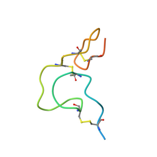
Coagulation factor IXB [auth D]50Homo sapiensMutation(s): 0 
Gene Names: F9
EC: 3.4.21.22
UniProt & NIH Common Fund Data Resources
Find proteins for P00740 (Homo sapiens)
Explore P00740 
Go to UniProtKB:  P00740
PHAROS:  P00740
GTEx:  ENSG00000101981 
Entity Groups  
Sequence Clusters30% Identity50% Identity70% Identity90% Identity95% Identity100% Identity
UniProt GroupP00740
Glycosylation
Glycosylation Sites: 1Go to GlyGen: P00740-1
Sequence Annotations
Expand
  • Reference Sequence
Oligosaccharides

Help

Entity ID: 3
MoleculeChains Length2D Diagram Glycosylation3D Interactions
alpha-D-xylopyranose-(1-3)-alpha-D-xylopyranose-(1-3)-beta-D-glucopyranoseC [auth B]3O-Glycosylation
Glycosylation Resources
GlyTouCan:  G41179DF
GlyCosmos:  G41179DF
GlyGen:  G41179DF
Small Molecules
Ligands 3 Unique
IDChains Name / Formula / InChI Key2D Diagram3D Interactions
UDP
Query on UDP

Download Ideal Coordinates CCD File 
E [auth A]URIDINE-5'-DIPHOSPHATE
C9 H14 N2 O12 P2
XCCTYIAWTASOJW-XVFCMESISA-N
SO4
Query on SO4

Download Ideal Coordinates CCD File 
F [auth A]SULFATE ION
O4 S
QAOWNCQODCNURD-UHFFFAOYSA-L
MN
Query on MN

Download Ideal Coordinates CCD File 
D [auth A]MANGANESE (II) ION
Mn
WAEMQWOKJMHJLA-UHFFFAOYSA-N
Experimental Data & Validation

Experimental Data

  • Method: X-RAY DIFFRACTION
  • Resolution: 2.08 Å
  • R-Value Free: 0.246 
  • R-Value Work: 0.191 
  • R-Value Observed: 0.194 
  • Space Group: P 3
Unit Cell:
Length ( Å )Angle ( ˚ )
a = 88.911α = 90
b = 88.911β = 90
c = 42.683γ = 120
Software Package:
Software NamePurpose
REFMACrefinement

Structure Validation

View Full Validation Report


Ligand Structure Quality Assessment 


Entry History 

Deposition Data

  • Released Date: 2015-09-30 
  • Deposition Author(s): Yu, H., Li, H.

Revision History  (Full details and data files)

  • Version 1.0: 2015-09-30
    Type: Initial release
  • Version 1.1: 2016-04-20
    Changes: Database references
  • Version 2.0: 2020-07-29
    Type: Remediation
    Reason: Carbohydrate remediation
    Changes: Atomic model, Data collection, Derived calculations, Non-polymer description, Structure summary
  • Version 2.1: 2023-12-27
    Changes: Data collection, Database references, Structure summary