


Applying Fragments Based- Drug Design to identify multiple binding modes on cysteine protease.
Tochowicz, A., Lee, G.M., Arkin, M.R., Neitz, J., McKerrow, J., Craik, C.S.To be published.
Experimental Data Snapshot
Starting Model: experimental
View more details
Entity ID: 1 | |||||
|---|---|---|---|---|---|
| Molecule | Chains | Sequence Length | Organism | Details | Image |
| Cruzipain | 216 | Trypanosoma cruzi | Mutation(s): 1 EC: 3.4.22.51 | 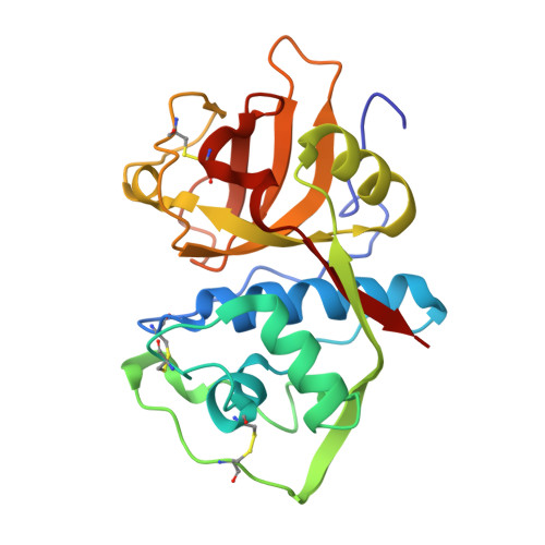 | |
UniProt | |||||
Entity Groups | |||||
| Sequence Clusters | 30% Identity50% Identity70% Identity90% Identity95% Identity100% Identity | ||||
| UniProt Group | P25779 | ||||
Sequence AnnotationsExpand | |||||
Reference Sequence | |||||
| Ligands 3 Unique | |||||
|---|---|---|---|---|---|
| ID | Chains | Name / Formula / InChI Key | 2D Diagram | 3D Interactions | |
| 3H6 Download:Ideal Coordinates CCD File | G [auth A] | N-(1H-benzimidazol-2-yl)-3-(4-fluorophenyl)-1H-pyrazole-4-carboxamide C17 H12 F N5 O NATFUWRFXYXTFK-UHFFFAOYSA-N |  | ||
| 3H5 Download:Ideal Coordinates CCD File | F [auth A], J [auth C] | N-(1H-benzimidazol-2-yl)-1,3-dimethyl-1H-pyrazole-4-carboxamide C13 H13 N5 O ATWRDFSNOOXDOA-UHFFFAOYSA-N |  | ||
| 3H7 Download:Ideal Coordinates CCD File | H [auth B], I [auth C], K [auth D] | 4,6-difluoro-1,3-benzothiazol-2-amine C7 H4 F2 N2 S DDKKXSCVPKDRRS-UHFFFAOYSA-N |  | ||
| Length ( Å ) | Angle ( ˚ ) |
|---|---|
| a = 137.79 | α = 90 |
| b = 137.79 | β = 90 |
| c = 166.48 | γ = 90 |
| Software Name | Purpose |
|---|---|
| PHENIX | refinement |
| MOSFLM | data reduction |
| XSCALE | data scaling |
| XDS | data reduction |
| SCALA | data scaling |


