4W4L | pdb_00004w4l

Crystal structure of EspG5 in complex with PE25 and PPE41 from the ESX-5 type VII secretion system of M. tuberculosis


Experimental Data Snapshot

  • Method: X-RAY DIFFRACTION
  • Resolution: 2.45 Å
  • R-Value Free: 
    0.230 (Depositor), 0.238 (DCC) 
  • R-Value Work: 
    0.202 (Depositor), 0.213 (DCC) 
  • R-Value Observed: 
    0.203 (Depositor) 

Starting Models: experimental
View more details

wwPDB Validation 3D Report Full Report

Validation slider image for 4W4L

This is version 1.5 of the entry. See complete history

Literature

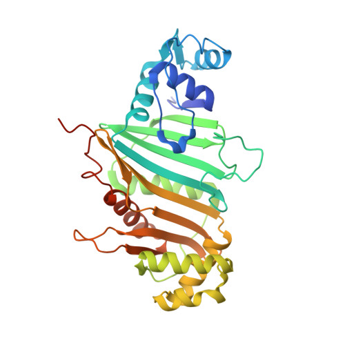
Structure of a PE-PPE-EspG complex from Mycobacterium tuberculosis reveals molecular specificity of ESX protein secretion.

Ekiert, D.C.Cox, J.S.

(2014) Proc Natl Acad Sci U S A 111: 14758-14763

  • DOI: https://doi.org/10.1073/pnas.1409345111
  • Primary Citation Related Structures: 
    4W4I, 4W4J, 4W4K, 4W4L

  • PubMed Abstract: 

    Nearly 10% of the coding capacity of the Mycobacterium tuberculosis genome is devoted to two highly expanded and enigmatic protein families called PE and PPE, some of which are important virulence/immunogenicity factors and are secreted during infection via a unique alternative secretory system termed "type VII." How PE-PPE proteins function during infection and how they are translocated to the bacterial surface through the five distinct type VII secretion systems [ESAT-6 secretion system (ESX)] of M. tuberculosis is poorly understood. Here, we report the crystal structure of a PE-PPE heterodimer bound to ESX secretion-associated protein G (EspG), which adopts a novel fold. This PE-PPE-EspG complex, along with structures of two additional EspGs, suggests that EspG acts as an adaptor that recognizes specific PE-PPE protein complexes via extensive interactions with PPE domains, and delivers them to ESX machinery for secretion. Surprisingly, secretion of most PE-PPE proteins in M. tuberculosis is likely mediated by EspG from the ESX-5 system, underscoring the importance of ESX-5 in mycobacterial pathogenesis. Moreover, our results indicate that PE-PPE domains function as cis-acting targeting sequences that are read out by EspGs, revealing the molecular specificity for secretion through distinct ESX pathways.


  • Organizational Affiliation
    • Department of Microbiology and Immunology, University of California, San Francisco, CA 94143 damian.ekiert@ucsf.edu jeffery.cox@ucsf.edu.

Macromolecule Content 

  • Total Structure Weight: 65.31 kDa 
  • Atom Count: 4,460 
  • Modeled Residue Count: 555 
  • Deposited Residue Count: 597 
  • Unique protein chains: 3

Macromolecules

Find similar proteins by:|  3D Structure
Entity ID: 1
MoleculeChains  Sequence LengthOrganismDetailsImage
PE family protein PE25109Mycobacterium tuberculosis str. Erdman = ATCC 35801Mutation(s): 0 
Gene Names: PE25ERDMAN_2675Q643_02517
Find similar proteins by:|  3D Structure
Entity ID: 2
MoleculeChains  Sequence LengthOrganismDetailsImage
PPE family protein PPE41174Mycobacterium tuberculosis str. Erdman = ATCC 35801Mutation(s): 0 
Gene Names: PPE41ERDMAN_2674Q643_02516
Find similar proteins by:|  3D Structure
Entity ID: 3
MoleculeChains  Sequence LengthOrganismDetailsImage
EspG5314Mycobacterium tuberculosis str. Erdman = ATCC 35801Mutation(s): 0 
Gene Names: ERDMAN_1984Q643_01851

Small Molecules

Ligands 1 Unique
IDChains Name / Formula / InChI Key2D Diagram3D Interactions
EDO

Query on EDO


Download:Ideal Coordinates CCD File
D [auth B]1,2-ETHANEDIOL
C2 H6 O2
LYCAIKOWRPUZTN-UHFFFAOYSA-N

Experimental Data & Validation

Experimental Data

  • Method: X-RAY DIFFRACTION
  • Resolution: 2.45 Å
  • R-Value Free:  0.230 (Depositor), 0.238 (DCC) 
  • R-Value Work:  0.202 (Depositor), 0.213 (DCC) 
  • R-Value Observed: 0.203 (Depositor) 
Space Group: P 61 2 2
Unit Cell:
Length ( Å )Angle ( ˚ )
a = 138.64α = 90
b = 138.64β = 90
c = 169.68γ = 120
Software Package:
Software NamePurpose
PHENIXrefinement
Blu-Icedata collection
XDSdata reduction
PHASERphasing

Structure Validation

View Full Validation Report


Entry History 

& Funding Information

Deposition Data


Funding OrganizationLocationGrant Number
National Institutes of Health/National Institute Of Allergy and Infectious Diseases (NIH/NIAID)United StatesR01AI081727
Damon Runyon Cancer Research FoundationUnited KingdomDRG-2140-12

Revision History  (Full details and data files)

  • Version 1.0: 2014-10-01
    Type: Initial release
  • Version 1.1: 2014-10-15
    Changes: Database references
  • Version 1.2: 2014-10-29
    Changes: Database references
  • Version 1.3: 2017-09-27
    Changes: Author supporting evidence, Database references, Derived calculations, Other, Refinement description, Source and taxonomy, Structure summary
  • Version 1.4: 2019-12-11
    Changes: Author supporting evidence
  • Version 1.5: 2023-09-27
    Changes: Data collection, Database references, Refinement description