4UWW | pdb_00004uww

Crystallographic Structure of the Intramineral Protein Struthicalcin from Struthio camelus Eggshell


Experimental Data Snapshot

  • Method: X-RAY DIFFRACTION
  • Resolution: 1.44 Å
  • R-Value Free: 
    0.232 (Depositor), 0.235 (DCC) 
  • R-Value Work: 
    0.213 (Depositor), 0.222 (DCC) 
  • R-Value Observed: 
    0.214 (Depositor) 

wwPDB Validation 3D Report Full Report

Validation slider image for 4UWW

This is version 1.2 of the entry. See complete history

Literature

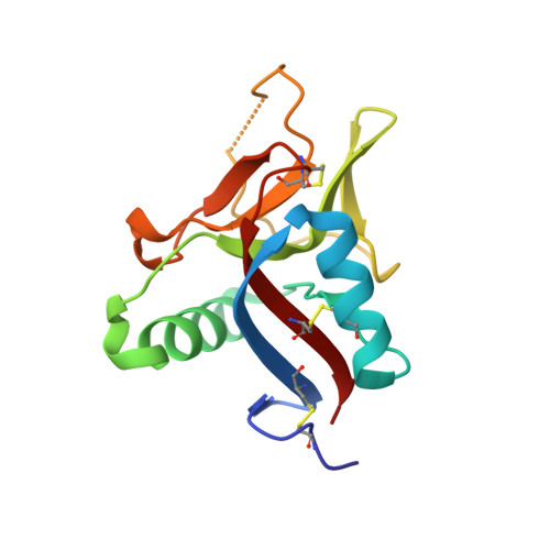
Crystal Structure of Struthiocalcin-1, an Intramineral Protein from Struthio Camelus Eggshell, in Two Different Crystal Forms.

Ruiz-Arellano, R.R.Medrano, F.J.Moreno, A.Romero, A.

(2015) Acta Crystallogr D Biol Crystallogr 71: 809

  • DOI: https://doi.org/10.1107/S139900471500125X
  • Primary Citation Related Structures: 
    4UWW, 4UXM

  • PubMed Abstract: 

    Biomineralization is the process by which living organisms produce minerals. One remarkable example is the formation of eggshells in birds. Struthiocalcins present in the ostrich (Struthio camellus) eggshell matrix act as biosensors of calcite growth during eggshell formation. Here, the crystal structure of struthiocalcin-1 (SCA-1) is reported in two different crystal forms. The structure is a compact single domain with an α/β fold characteristic of the C-type lectin family. In contrast to the related avian ovocleidin OC17, the electrostatic potential on the molecular surface is dominated by an acidic patch. Scanning electron microscopy combined with Raman spectroscopy indicates that these intramineral proteins (SCA-1 and SCA-2) induce calcium carbonate precipitation, leading to the formation of a stable form of calcite in the mature eggshell. Finally, the implications of these two intramineral proteins SCA-1 and SCA-2 in the nucleation of calcite during the formation of eggshells in ratite birds are discussed.


  • Organizational Affiliation
    • Instituto de Química, Universidad Nacional Autónoma de México, 04510 Mexico City, Mexico.

Macromolecule Content 

  • Total Structure Weight: 15.38 kDa 
  • Atom Count: 1,153 
  • Modeled Residue Count: 130 
  • Deposited Residue Count: 132 
  • Unique protein chains: 1

Macromolecules

Find similar proteins by:|  3D Structure
Entity ID: 1
MoleculeChains  Sequence LengthOrganismDetailsImage
STRUTHIOCALCIN-1132Struthio camelusMutation(s): 0 
UniProt
Find proteins for P83514 (Struthio camelus)
Explore P83514 
Go to UniProtKB:  P83514
Entity Groups
Sequence Clusters30% Identity50% Identity70% Identity90% Identity95% Identity100% Identity
UniProt GroupP83514
Sequence Annotations
Expand
Reference Sequence

Experimental Data & Validation

Experimental Data

  • Method: X-RAY DIFFRACTION
  • Resolution: 1.44 Å
  • R-Value Free:  0.232 (Depositor), 0.235 (DCC) 
  • R-Value Work:  0.213 (Depositor), 0.222 (DCC) 
  • R-Value Observed: 0.214 (Depositor) 
Space Group: P 21 21 2
Unit Cell:
Length ( Å )Angle ( ˚ )
a = 55.94α = 90
b = 72.6β = 90
c = 37.1γ = 90
Software Package:
Software NamePurpose
REFMACrefinement
MOSFLMdata reduction
SCALAdata scaling
PHASERphasing

Structure Validation

View Full Validation Report


Entry History 

Deposition Data

Revision History  (Full details and data files)

  • Version 1.0: 2015-04-08
    Type: Initial release
  • Version 1.1: 2015-04-22
    Changes: Database references
  • Version 1.2: 2024-10-23
    Changes: Data collection, Database references, Other, Structure summary