4UW0 | pdb_00004uw0

Low resolution structure of WbdD with C-terminal bundle ordered to residue 505


Experimental Data Snapshot

  • Method: X-RAY DIFFRACTION
  • Resolution: 3.87 Å
  • R-Value Free: 
    0.341 (Depositor), 0.353 (DCC) 
  • R-Value Work: 
    0.268 (Depositor), 0.277 (DCC) 
  • R-Value Observed: 
    0.271 (Depositor) 

wwPDB Validation 3D Report Full Report

Validation slider image for 4UW0

Ligand Structure Quality Assessment 


This is version 1.6 of the entry. See complete history. 

Literature

A Coiled-Coil Domain Acts as a Molecular Ruler to Regulate O-Antigen Chain Length in Lipopolysaccharide.

Hagelueken, G., Clarke, B.R., Huang, H., Tuukkanen, A., Danciu, I., Svergun, D.I., Hussain, R., Liu, H., Whitfield, C., Naismith, J.H.

(2015) Nat Struct Mol Biol 22: 50

  • DOI: https://doi.org/10.1038/nsmb.2935
  • Primary Citation Related Structures: 
    4UW0

  • PubMed Abstract: 

    Long-chain bacterial polysaccharides have important roles in pathogenicity. In Escherichia coli O9a, a model for ABC transporter-dependent polysaccharide assembly, a large extracellular carbohydrate with a narrow size distribution is polymerized from monosaccharides by a complex of two proteins, WbdA (polymerase) and WbdD (terminating protein). Combining crystallography and small-angle X-ray scattering, we found that the C-terminal domain of WbdD contains an extended coiled-coil that physically separates WbdA from the catalytic domain of WbdD. The effects of insertions and deletions in the coiled-coil region were analyzed in vivo, revealing that polymer size is controlled by varying the length of the coiled-coil domain. Thus, the coiled-coil domain of WbdD functions as a molecular ruler that, along with WbdA:WbdD stoichiometry, controls the chain length of a model bacterial polysaccharide.


  • Organizational Affiliation: 
    • Biomedical Sciences Research Complex, University of St Andrews, St Andrews, Fife, KY16 9ST, UK.

Macromolecule Content 

  • Total Structure Weight: 58.14 kDa 
  • Atom Count: 4,074 
  • Modeled Residue Count: 501 
  • Deposited Residue Count: 505 
  • Unique protein chains: 1

Macromolecules

Find similar proteins by:|  3D Structure
Entity ID: 1
MoleculeChains  Sequence LengthOrganismDetailsImage
WBDD505Escherichia coliMutation(s): 0 
Gene Names: EC3426_03072
EC: 2.1.1.294 (UniProt), 2.7.1.181 (UniProt)
UniProt
Find proteins for J7I4B7 (Escherichia coli)
Explore J7I4B7 
Go to UniProtKB:  J7I4B7
Entity Groups
Sequence Clusters30% Identity50% Identity70% Identity90% Identity95% Identity100% Identity
UniProt GroupJ7I4B7
Sequence Annotations
Expand
Reference Sequence

Small Molecules

Ligands 1 Unique
IDChains Name / Formula / InChI Key2D Diagram3D Interactions
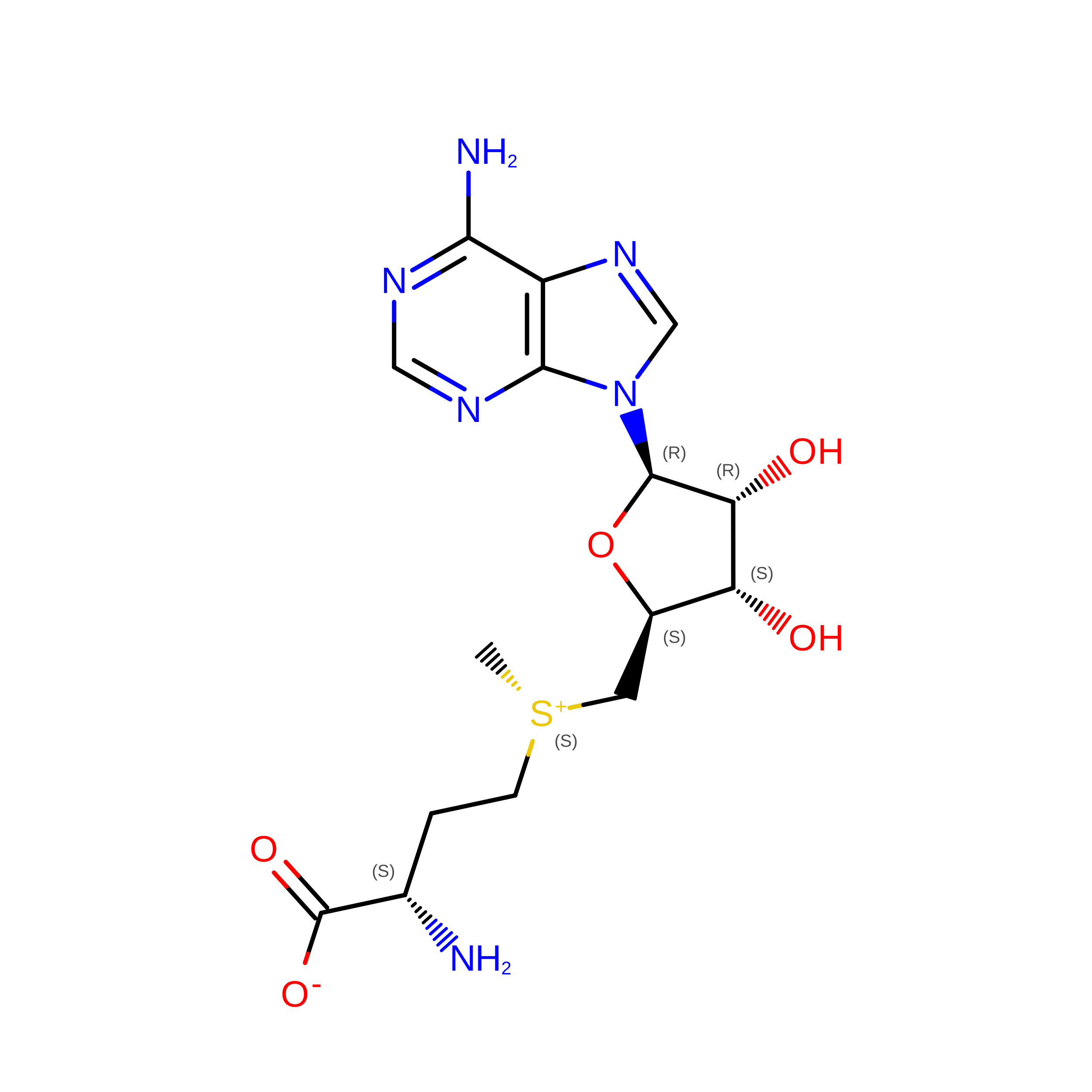
SAM

Query on SAM


Download:Ideal Coordinates CCD File
B [auth A]S-ADENOSYLMETHIONINE
C15 H22 N6 O5 S
MEFKEPWMEQBLKI-FCKMPRQPSA-N

Experimental Data & Validation

Experimental Data

  • Method: X-RAY DIFFRACTION
  • Resolution: 3.87 Å
  • R-Value Free:  0.341 (Depositor), 0.353 (DCC) 
  • R-Value Work:  0.268 (Depositor), 0.277 (DCC) 
  • R-Value Observed: 0.271 (Depositor) 
Space Group: I 2 3
Unit Cell:
Length ( Å )Angle ( ˚ )
a = 181.29α = 90
b = 181.29β = 90
c = 181.29γ = 90
Software Package:
Software NamePurpose
REFMACrefinement

Structure Validation

View Full Validation Report


Ligand Structure Quality Assessment 


Entry History 

Deposition Data

Revision History  (Full details and data files)

  • Version 1.0: 2014-08-27
    Type: Initial release
  • Version 1.1: 2014-12-10
    Changes: Database references
  • Version 1.2: 2014-12-31
    Changes: Database references
  • Version 1.3: 2015-01-21
    Changes: Database references
  • Version 1.4: 2018-04-25
    Changes: Advisory, Data collection
  • Version 1.5: 2019-09-18
    Changes: Data collection, Database references, Source and taxonomy
  • Version 1.6: 2024-05-08
    Changes: Advisory, Data collection, Database references, Derived calculations, Other