Laboratory Evolved Enzymes Provide Snapshots of the Development of Enantioconvergence in Enzyme-Catalyzed Epoxide Hydrolysis.
Janfalk Carlsson, A., Bauer, P., Dobritzsch, D., Nilsson, M., Kamerlin, S.C., Widersten, M.(2016) Chembiochem 17: 1693
- PubMed: 27383542
- DOI: https://doi.org/10.1002/cbic.201600330
- Primary Citation of Related Structures:
4UFO, 4UFP, 4UHB - PubMed Abstract:
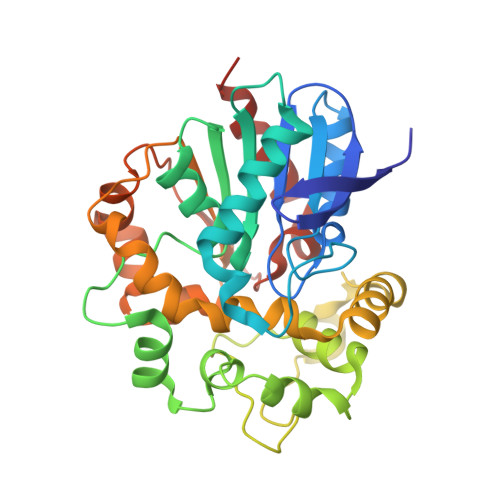
Engineered enzyme variants of potato epoxide hydrolase (StEH1) display varying degrees of enrichment of (2R)-3-phenylpropane-1,2-diol from racemic benzyloxirane. Curiously, the observed increase in the enantiomeric excess of the (R)-diol is not only a consequence of changes in enantioselectivity for the preferred epoxide enantiomer, but also to changes in the regioselectivity of the epoxide ring opening of (S)-benzyloxirane. In order to probe the structural origin of these differences in substrate selectivity and catalytic regiopreference, we solved the crystal structures for the evolved StEH1 variants. We used these structures as a starting point for molecular docking studies of the epoxide enantiomers into the respective active sites. Interestingly, despite the simplicity of our docking analysis, the apparent preferred binding modes appear to rationalize the experimentally determined regioselectivities. The analysis also identifies an active site residue (F33) as a potentially important interaction partner, a role that could explain the high conservation of this residue during evolution. Overall, our experimental, structural, and computational studies provide snapshots into the evolution of enantioconvergence in StEH1-catalyzed epoxide hydrolysis.
- Department of Chemistry, BMC, Uppsala University, Box 576, 751 23, Uppsala, Sweden.
Organizational Affiliation:


