4TQJ | pdb_00004tqj

Structural basis of specific recognition of non-reducing terminal N-acetylglucosamine by an Agrocybe aegerita lection


Experimental Data Snapshot

  • Method: X-RAY DIFFRACTION
  • Resolution: 2.00 Å
  • R-Value Free: 
    0.235 (Depositor), 0.228 (DCC) 
  • R-Value Work: 
    0.187 (Depositor), 0.183 (DCC) 
  • R-Value Observed: 
    0.190 (Depositor) 

wwPDB Validation 3D Report Full Report

Validation slider image for 4TQJ

This is version 1.2 of the entry. See complete history

Literature

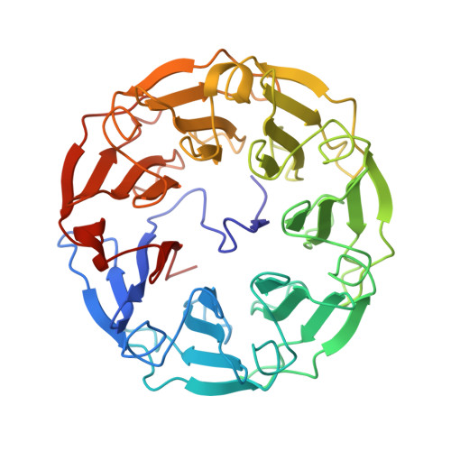
Structural Basis of Specific Recognition of Non-Reducing Terminal N-Acetylglucosamine by an Agrocybe aegerita Lectin.

Ren, X.M.Li, D.F.Jiang, S.Lan, X.Q.Hu, Y.Sun, H.Wang, D.C.

(2015) PLoS One 10: e0129608-e0129608

  • DOI: https://doi.org/10.1371/journal.pone.0129608
  • Primary Citation Related Structures: 
    4TQJ, 4TQK, 4TQM

  • PubMed Abstract: 

    O-linked N-acetylglucosaminylation (O-GlcNAcylation) is a reversible post-translational modification that plays essential roles in many cellular pathways. Research in this field, however, is hampered by the lack of suitable probes to identify, accumulate, and purify the O-GlcNAcylated proteins. We have previously reported the identification of a lectin from the mushroom Agrocybe aegerita, i.e., Agrocybe aegerita lectin 2, or AAL2, that could bind terminal N-acetylglucosamine with higher affinities and specificity than other currently used probes. In this paper, we report the crystal structures of AAL2 and its complexes with GlcNAc and GlcNAcβ1-3Galβ1-4GlcNAc and reveal the structural basis of GlcNAc recognition by AAL2 and residues essential for the binding of terminal N-acetylglucosamine. Study on AAL2 may enable us to design a protein probe that can be used to identify and purify O-GlcNAcylated proteins more efficiently.


  • Organizational Affiliation
    • National Laboratory of Biomacromolecules, Institute of Biophysics, Chinese Academy of Sciences, 15 Datun Road, Beijing, 100101, People's Republic of China; University of Chinese Academy of Sciences, No. 19A Yuquan Road, Beijing, 100049, People's Republic of China.

Macromolecule Content 

  • Total Structure Weight: 86.16 kDa 
  • Atom Count: 6,842 
  • Modeled Residue Count: 806 
  • Deposited Residue Count: 812 
  • Unique protein chains: 1

Macromolecules

Find similar proteins by:|  3D Structure
Entity ID: 1
MoleculeChains  Sequence LengthOrganismDetailsImage
Lectin 2
A, B
406Cyclocybe aegeritaMutation(s): 1 
Gene Names: aal-2
UniProt
Find proteins for H6CS64 (Cyclocybe aegerita)
Explore H6CS64 
Go to UniProtKB:  H6CS64
Entity Groups
Sequence Clusters30% Identity50% Identity70% Identity90% Identity95% Identity100% Identity
UniProt GroupH6CS64
Sequence Annotations
Expand
Reference Sequence

Experimental Data & Validation

Experimental Data

  • Method: X-RAY DIFFRACTION
  • Resolution: 2.00 Å
  • R-Value Free:  0.235 (Depositor), 0.228 (DCC) 
  • R-Value Work:  0.187 (Depositor), 0.183 (DCC) 
  • R-Value Observed: 0.190 (Depositor) 
Space Group: P 21 21 21
Unit Cell:
Length ( Å )Angle ( ˚ )
a = 52.6α = 90
b = 111.7β = 90
c = 135.97γ = 90
Software Package:
Software NamePurpose
iMOSFLMdata reduction
PDB_EXTRACTdata extraction
PHENIXrefinement
MOSFLMdata reduction
SCALAdata scaling

Structure Validation

View Full Validation Report


Entry History 

Deposition Data

Revision History  (Full details and data files)

  • Version 1.0: 2015-06-03
    Type: Initial release
  • Version 1.1: 2015-08-05
    Changes: Database references
  • Version 1.2: 2024-03-20
    Changes: Data collection, Database references, Derived calculations