4RG5 | pdb_00004rg5

Crystal Structure of S. Pombe SMN YG-Dimer


Experimental Data Snapshot

  • Method: X-RAY DIFFRACTION
  • Resolution: 1.70 Å
  • R-Value Free: 
    0.192 (Depositor), 0.192 (DCC) 
  • R-Value Work: 
    0.165 (Depositor), 0.165 (DCC) 
  • R-Value Observed: 
    0.166 (Depositor) 

wwPDB Validation 3D Report Full Report

Validation slider image for 4RG5

This is version 2.1 of the entry. See complete history

Literature

Oligomeric Properties of Survival Motor NeuronGemin2 Complexes.

Gupta, K.Martin, R.Sharp, R.Sarachan, K.L.Ninan, N.S.Van Duyne, G.D.

(2015) J Biological Chem 290: 20185-20199

  • DOI: https://doi.org/10.1074/jbc.M115.667279
  • Primary Citation Related Structures: 
    4RG5

  • PubMed Abstract: 

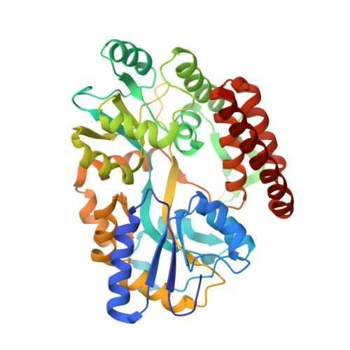
    The survival motor neuron (SMN) protein forms the oligomeric core of a multiprotein complex required for the assembly of spliceosomal small nuclear ribonucleoproteins. Deletions and mutations in the SMN1 gene are associated with spinal muscular atrophy (SMA), a devastating neurodegenerative disease that is the leading heritable cause of infant mortality. Oligomerization of SMN is required for its function, and some SMA patient mutations disrupt the ability of SMN to self-associate. Here, we investigate the oligomeric nature of the SMN·Gemin2 complexes from humans and fission yeast (hSMN·Gemin2 and ySMN·Gemin2). We find that hSMN·Gemin2 forms oligomers spanning the dimer to octamer range. The YG box oligomerization domain of SMN is both necessary and sufficient to form these oligomers. ySMN·Gemin2 exists as a dimer-tetramer equilibrium with Kd = 1.0 ± 0.9 μM. A 1.9 Å crystal structure of the ySMN YG box confirms a high level of structural conservation with the human ortholog in this important region of SMN. Disulfide cross-linking experiments indicate that SMN tetramers are formed by self-association of stable, non-dissociating dimers. Thus, SMN tetramers do not form symmetric helical bundles such as those found in glycine zipper transmembrane oligomers. The dimer-tetramer nature of SMN complexes and the dimer of dimers organization of the SMN tetramer provide an important foundation for ongoing studies to understand the mechanism of SMN-assisted small nuclear ribonucleoprotein assembly and the underlying causes of SMA.


  • Organizational Affiliation
    • From the Department of Biochemistry and Biophysics and.

Macromolecule Content 

  • Total Structure Weight: 90.38 kDa 
  • Atom Count: 7,253 
  • Modeled Residue Count: 801 
  • Deposited Residue Count: 802 
  • Unique protein chains: 1

Macromolecules

Find similar proteins by:|  3D Structure
Entity ID: 1
MoleculeChains  Sequence LengthOrganismDetailsImage
Maltose-binding periplasmic protein, Survival Motor Neuron protein chimera
A, B
401Escherichia coliSchizosaccharomyces pombe 972h-
This entity is chimeric
Mutation(s): 0 
UniProt
Find proteins for P0AEX9 (Escherichia coli (strain K12))
Explore P0AEX9 
Go to UniProtKB:  P0AEX9
Find proteins for Q09808 (Schizosaccharomyces pombe (strain 972 / ATCC 24843))
Explore Q09808 
Go to UniProtKB:  Q09808
Entity Groups
Sequence Clusters30% Identity50% Identity70% Identity90% Identity95% Identity100% Identity
UniProt GroupsQ09808P0AEX9
Sequence Annotations
Expand
Reference Sequence

Oligosaccharides

Help  
Entity ID: 2
MoleculeChains Length2D Diagram GlycosylationD Interactions
alpha-D-glucopyranose-(1-4)-alpha-D-glucopyranose
C, D
2N/A
Glycosylation Resources
GlyTouCan: G07411ON
GlyCosmos: G07411ON

Small Molecules

Ligands 2 Unique
IDChains Name / Formula / InChI Key2D Diagram3D Interactions
MLI

Query on MLI


Download:Ideal Coordinates CCD File
L [auth B],
M [auth B]
MALONATE ION
C3 H2 O4
OFOBLEOULBTSOW-UHFFFAOYSA-L
SO4

Query on SO4


Download:Ideal Coordinates CCD File
E [auth A]
F [auth A]
G [auth A]
H [auth A]
I [auth A]
E [auth A],
F [auth A],
G [auth A],
H [auth A],
I [auth A],
J [auth A],
K [auth B]
SULFATE ION
O4 S
QAOWNCQODCNURD-UHFFFAOYSA-L

Biologically Interesting Molecules (External Reference) 

1 Unique

Experimental Data & Validation

Experimental Data

  • Method: X-RAY DIFFRACTION
  • Resolution: 1.70 Å
  • R-Value Free:  0.192 (Depositor), 0.192 (DCC) 
  • R-Value Work:  0.165 (Depositor), 0.165 (DCC) 
  • R-Value Observed: 0.166 (Depositor) 
Space Group: P 21 21 21
Unit Cell:
Length ( Å )Angle ( ˚ )
a = 96.429α = 90
b = 105.015β = 90
c = 107.686γ = 90
Software Package:
Software NamePurpose
HKL-3000data collection
AMoREphasing
REFMACrefinement
HKL-3000data reduction
HKL-3000data scaling

Structure Validation

View Full Validation Report


Entry History 

Deposition Data

Revision History  (Full details and data files)

  • Version 1.0: 2015-07-01
    Type: Initial release
  • Version 1.1: 2015-09-16
    Changes: Database references
  • Version 1.2: 2017-08-02
    Changes: Source and taxonomy
  • Version 2.0: 2020-07-29
    Type: Remediation
    Reason: Carbohydrate remediation
    Changes: Atomic model, Data collection, Derived calculations, Non-polymer description, Structure summary
  • Version 2.1: 2024-02-28
    Changes: Data collection, Database references, Structure summary