4R2G | pdb_00004r2g

Crystal Structure of PGT124 Fab bound to HIV-1 JRCSF gp120 core and to CD4


Experimental Data Snapshot

  • Method: X-RAY DIFFRACTION
  • Resolution: 3.28 Å
  • R-Value Free: 
    0.263 (Depositor), 0.266 (DCC) 
  • R-Value Work: 
    0.207 (Depositor), 0.211 (DCC) 
  • R-Value Observed: 
    0.210 (Depositor) 

wwPDB Validation 3D Report Full Report

Validation slider image for 4R2G

Ligand Structure Quality Assessment 


This is version 3.0 of the entry. See complete history

Literature

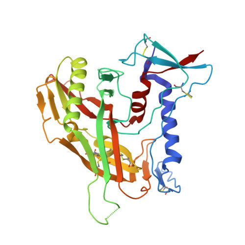
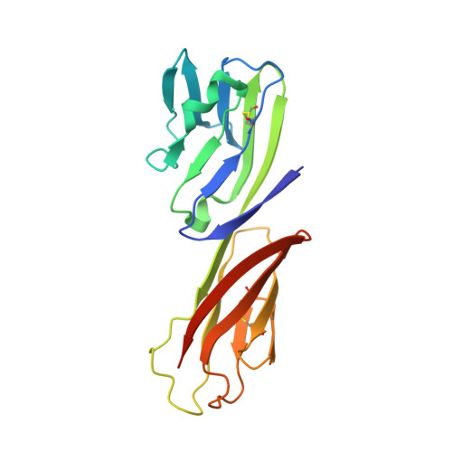
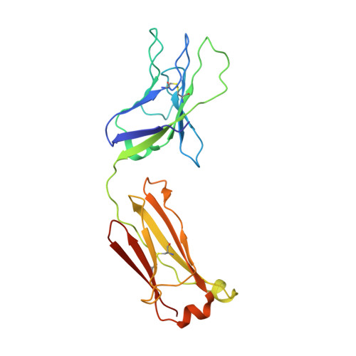
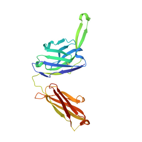
Structural Evolution of Glycan Recognition by a Family of Potent HIV Antibodies.

Garces, F.Sok, D.Kong, L.McBride, R.Kim, H.J.Saye-Francisco, K.F.Julien, J.P.Hua, Y.Cupo, A.Moore, J.P.Paulson, J.C.Ward, A.B.Burton, D.R.Wilson, I.A.

(2014) Cell 159: 69-79

  • DOI: https://doi.org/10.1016/j.cell.2014.09.009
  • Primary Citation Related Structures: 
    4R26, 4R2G

  • PubMed Abstract: 

    The HIV envelope glycoprotein (Env) is densely covered with self-glycans that should help shield it from recognition by the human immune system. Here, we examine how a particularly potent family of broadly neutralizing antibodies (Abs) has evolved common and distinct structural features to counter the glycan shield and interact with both glycan and protein components of HIV Env. The inferred germline antibody already harbors potential binding pockets for a glycan and a short protein segment. Affinity maturation then leads to divergent evolutionary branches that either focus on a single glycan and protein segment (e.g., Ab PGT124) or engage multiple glycans (e.g., Abs PGT121-123). Furthermore, other surrounding glycans are avoided by selecting an appropriate initial antibody shape that prevents steric hindrance. Such molecular recognition lessons are important for engineering proteins that can recognize or accommodate glycans.


  • Organizational Affiliation
    • Department of Integrative Structural and Computational Biology, The Scripps Research Institute, La Jolla, CA 92037, USA; International AIDS Vaccine Initiative Neutralizing Antibody Center, The Scripps Research Institute, La Jolla, CA 92037, USA; Scripps Center for HIV/AIDS Vaccine Immunology & Immunogen Discovery, The Scripps Research Institute, La Jolla, CA 92037, USA.

Macromolecule Content 

  • Total Structure Weight: 428.76 kDa 
  • Atom Count: 29,158 
  • Modeled Residue Count: 3,657 
  • Deposited Residue Count: 3,772 
  • Unique protein chains: 4

Macromolecules

Find similar proteins by:|  3D Structure
Entity ID: 1
MoleculeChains  Sequence LengthOrganismDetailsImage
Surface protein gp160A [auth E],
N [auth O],
O [auth A],
P [auth K]
309Human immunodeficiency virus type 1 (JRCSF ISOLATE)Mutation(s): 0 
Gene Names: env
UniProt
Find proteins for P20871 (Human immunodeficiency virus type 1 group M subtype B (isolate JRCSF))
Explore P20871 
Go to UniProtKB:  P20871
Entity Groups
Sequence Clusters30% Identity50% Identity70% Identity90% Identity95% Identity100% Identity
UniProt GroupP20871
Glycosylation
Glycosylation Sites: 6
Sequence Annotations
Expand
Reference Sequence
Find similar proteins by:|  3D Structure
Entity ID: 2
MoleculeChains  Sequence LengthOrganismDetailsImage
T-cell surface glycoprotein CD4B [auth F],
E [auth B],
H,
K [auth L]
184Homo sapiensMutation(s): 0 
Gene Names: CD4
UniProt & NIH Common Fund Data Resources
Find proteins for P01730 (Homo sapiens)
Explore P01730 
Go to UniProtKB:  P01730
PHAROS:  P01730
GTEx:  ENSG00000010610 
Entity Groups
Sequence Clusters30% Identity50% Identity70% Identity90% Identity95% Identity100% Identity
UniProt GroupP01730
Sequence Annotations
Expand
Reference Sequence
Find similar proteins by:|  3D Structure
Entity ID: 3
MoleculeChains  Sequence LengthOrganismDetailsImage
PGT124 Light ChainC [auth P],
F [auth C],
I,
L [auth M]
214Homo sapiensMutation(s): 0 
Gene Names: IGL@IGLC3
UniProt & NIH Common Fund Data Resources
Find proteins for P0DOY3 (Homo sapiens)
Explore P0DOY3 
Go to UniProtKB:  P0DOY3
GTEx:  ENSG00000211679 
Entity Groups
Sequence Clusters30% Identity50% Identity70% Identity90% Identity95% Identity100% Identity
UniProt GroupP0DOY3
Sequence Annotations
Expand
Reference Sequence
Find similar proteins by:|  3D Structure
Entity ID: 4
MoleculeChains  Sequence LengthOrganismDetailsImage
PGT124 Heavy ChainD [auth Q],
G [auth D],
J,
M [auth N]
236Homo sapiensMutation(s): 0 
Gene Names: IGH@
UniProt
Find proteins for P0DOX5 (Homo sapiens)
Explore P0DOX5 
Go to UniProtKB:  P0DOX5
Entity Groups
Sequence Clusters30% Identity50% Identity70% Identity90% Identity95% Identity100% Identity
UniProt GroupP0DOX5
Sequence Annotations
Expand
Reference Sequence

Oligosaccharides

Help  
Entity ID: 5
MoleculeChains Length2D Diagram GlycosylationD Interactions
alpha-D-mannopyranose-(1-2)-alpha-D-mannopyranose-(1-2)-alpha-D-mannopyranose-(1-3)-[alpha-D-mannopyranose-(1-2)-alpha-D-mannopyranose-(1-6)-[alpha-D-mannopyranose-(1-3)]alpha-D-mannopyranose-(1-6)]beta-D-mannopyranose-(1-4)-2-acetamido-2-deoxy-beta-D-glucopyranose-(1-4)-2-acetamido-2-deoxy-beta-D-glucopyranoseQ [auth G],
R,
S,
V
10N-Glycosylation
Glycosylation Resources
GlyTouCan: G40702WU
GlyCosmos: G40702WU
GlyGen: G40702WU
Entity ID: 6
MoleculeChains Length2D Diagram GlycosylationD Interactions
2-acetamido-2-deoxy-beta-D-glucopyranose-(1-4)-2-acetamido-2-deoxy-beta-D-glucopyranose
T, U
2N-Glycosylation
Glycosylation Resources
GlyTouCan: G42666HT
GlyCosmos: G42666HT
GlyGen: G42666HT

Small Molecules

Ligands 3 Unique
IDChains Name / Formula / InChI Key2D Diagram3D Interactions
NAG

Query on NAG


Download:Ideal Coordinates CCD File
AA [auth E]
AB [auth K]
BA [auth E]
BB [auth K]
CA [auth E]
AA [auth E],
AB [auth K],
BA [auth E],
BB [auth K],
CA [auth E],
CB [auth K],
DA [auth E],
JA [auth O],
KA [auth O],
LA [auth O],
MA [auth O],
NA [auth O],
OA [auth O],
PA [auth O],
RA [auth A],
SA [auth A],
TA [auth A],
UA [auth A],
VA [auth A],
W [auth E],
WA [auth A],
X [auth E],
Y [auth E],
YA [auth K],
Z [auth E],
ZA [auth K]
2-acetamido-2-deoxy-beta-D-glucopyranose
C8 H15 N O6
OVRNDRQMDRJTHS-FMDGEEDCSA-N
GOL

Query on GOL


Download:Ideal Coordinates CCD File
FA [auth Q],
GA [auth D],
HA [auth J],
IA [auth N]
GLYCEROL
C3 H8 O3
PEDCQBHIVMGVHV-UHFFFAOYSA-N
CL

Query on CL


Download:Ideal Coordinates CCD File
DB [auth K],
EA [auth E],
QA [auth O],
XA [auth A]
CHLORIDE ION
Cl
VEXZGXHMUGYJMC-UHFFFAOYSA-M

Experimental Data & Validation

Experimental Data

  • Method: X-RAY DIFFRACTION
  • Resolution: 3.28 Å
  • R-Value Free:  0.263 (Depositor), 0.266 (DCC) 
  • R-Value Work:  0.207 (Depositor), 0.211 (DCC) 
  • R-Value Observed: 0.210 (Depositor) 
Space Group: P 21 21 21
Unit Cell:
Length ( Å )Angle ( ˚ )
a = 164.406α = 90
b = 165.438β = 90
c = 229.712γ = 90
Software Package:
Software NamePurpose
HKL-2000data collection
PHASERphasing
PHENIXrefinement
HKL-2000data reduction
HKL-2000data scaling

Structure Validation

View Full Validation Report


Ligand Structure Quality Assessment 


Entry History 

Deposition Data

Revision History  (Full details and data files)

  • Version 1.0: 2014-10-08
    Type: Initial release
  • Version 1.1: 2014-10-15
    Changes: Derived calculations
  • Version 1.2: 2017-06-21
    Changes: Database references, Source and taxonomy, Structure summary
  • Version 2.0: 2020-07-29
    Type: Remediation
    Reason: Carbohydrate remediation
    Changes: Advisory, Atomic model, Data collection, Derived calculations, Structure summary
  • Version 3.0: 2024-12-25
    Changes: Advisory, Atomic model, Data collection, Database references, Derived calculations, Structure summary