4QMH | pdb_00004qmh

The XMAP215 family drives microtubule polymerization using a structurally diverse TOG array


Experimental Data Snapshot

  • Method: X-RAY DIFFRACTION
  • Resolution: 1.65 Å
  • R-Value Free: 
    0.187 (Depositor), 0.186 (DCC) 
  • R-Value Work: 
    0.157 (Depositor), 0.155 (DCC) 
  • R-Value Observed: 
    0.159 (Depositor) 

wwPDB Validation 3D Report Full Report

Validation slider image for 4QMH

This is version 1.2 of the entry. See complete history

Literature

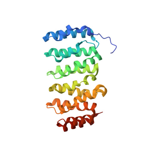
The XMAP215 family drives microtubule polymerization using a structurally diverse TOG array.

Fox, J.C.Howard, A.E.Currie, J.D.Rogers, S.L.Slep, K.C.

(2014) Mol Biol Cell 25: 2375-2392

  • DOI: https://doi.org/10.1091/mbc.E13-08-0501
  • Primary Citation Related Structures: 
    4QMH, 4QMI, 4QMJ

  • PubMed Abstract: 

    XMAP215 family members are potent microtubule (MT) polymerases, with mutants displaying reduced MT growth rates and aberrant spindle morphologies. XMAP215 proteins contain arrayed tumor overexpressed gene (TOG) domains that bind tubulin. Whether these TOG domains are architecturally equivalent is unknown. Here we present crystal structures of TOG4 from Drosophila Msps and human ch-TOG. These TOG4 structures architecturally depart from the structures of TOG domains 1 and 2, revealing a conserved domain bend that predicts a novel engagement with α-tubulin. In vitro assays show differential tubulin-binding affinities across the TOG array, as well as differential effects on MT polymerization. We used Drosophila S2 cells depleted of endogenous Msps to assess the importance of individual TOG domains. Whereas a TOG1-4 array largely rescues MT polymerization rates, mutating tubulin-binding determinants in any single TOG domain dramatically reduces rescue activity. Our work highlights the structurally diverse yet positionally conserved TOG array that drives MT polymerization.


  • Organizational Affiliation
    • Department of Biochemistry and Biophysics, University of North Carolina at Chapel Hill, Chapel Hill, NC 27599Graduate Program in Molecular and Cellular Biophysics, University of North Carolina at Chapel Hill, Chapel Hill, NC 27599.

Macromolecule Content 

  • Total Structure Weight: 26.4 kDa 
  • Atom Count: 2,211 
  • Modeled Residue Count: 236 
  • Deposited Residue Count: 241 
  • Unique protein chains: 1

Macromolecules

Find similar proteins by:|  3D Structure
Entity ID: 1
MoleculeChains  Sequence LengthOrganismDetailsImage
LP04448p241Drosophila melanogasterMutation(s): 0 
Gene Names: CG5000CG5000 Dmel_CG5000Dmel_CG5000msps
UniProt
Find proteins for Q9VEZ3 (Drosophila melanogaster)
Explore Q9VEZ3 
Go to UniProtKB:  Q9VEZ3
Entity Groups
Sequence Clusters30% Identity50% Identity70% Identity90% Identity95% Identity100% Identity
UniProt GroupQ9VEZ3
Sequence Annotations
Expand
Reference Sequence

Small Molecules

Ligands 1 Unique
IDChains Name / Formula / InChI Key2D Diagram3D Interactions
SO4

Query on SO4


Download:Ideal Coordinates CCD File
B [auth A]SULFATE ION
O4 S
QAOWNCQODCNURD-UHFFFAOYSA-L

Experimental Data & Validation

Experimental Data

  • Method: X-RAY DIFFRACTION
  • Resolution: 1.65 Å
  • R-Value Free:  0.187 (Depositor), 0.186 (DCC) 
  • R-Value Work:  0.157 (Depositor), 0.155 (DCC) 
  • R-Value Observed: 0.159 (Depositor) 
Space Group: P 1
Unit Cell:
Length ( Å )Angle ( ˚ )
a = 31.788α = 100.05
b = 32.613β = 95.23
c = 59.795γ = 109.95
Software Package:
Software NamePurpose
ADSCdata collection
PHENIXmodel building
PHENIXrefinement
HKL-2000data reduction
HKL-2000data scaling
PHENIXphasing

Structure Validation

View Full Validation Report


Entry History 

Deposition Data

Revision History  (Full details and data files)

  • Version 1.0: 2014-07-09
    Type: Initial release
  • Version 1.1: 2014-09-03
    Changes: Database references
  • Version 1.2: 2024-02-28
    Changes: Data collection, Database references, Derived calculations