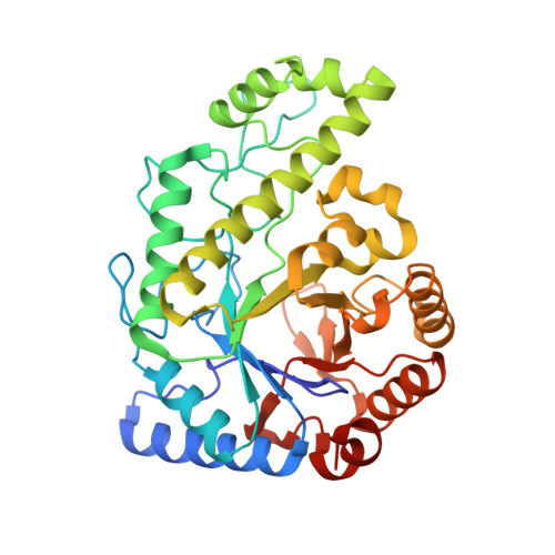
4P7L

Structure of Escherichia coli PgaB C-terminal domain, P212121 crystal form


Experimental Data Snapshot

  • Method: X-RAY DIFFRACTION
  • Resolution: 1.80 Å
  • R-Value Free: 0.189 
  • R-Value Work: 0.142 
  • R-Value Observed: 0.145 

Starting Model: experimental
View more details

wwPDB Validation   3D Report Full Report


This is version 1.6 of the entry. See complete history


Literature

Modification and periplasmic translocation of the biofilm exopolysaccharide poly-beta-1,6-N-acetyl-D-glucosamine.

Little, D.J.Li, G.Ing, C.DiFrancesco, B.R.Bamford, N.C.Robinson, H.Nitz, M.Pomes, R.Howell, P.L.

(2014) Proc Natl Acad Sci U S A 111: 11013-11018

  • DOI: https://doi.org/10.1073/pnas.1406388111
  • Primary Citation of Related Structures:  
    4P7L, 4P7N, 4P7O, 4P7Q, 4P7R

  • PubMed Abstract: 

    Poly-β-1,6-N-acetyl-D-glucosamine (PNAG) is an exopolysaccharide produced by a wide variety of medically important bacteria. Polyglucosamine subunit B (PgaB) is responsible for the de-N-acetylation of PNAG, a process required for polymer export and biofilm formation. PgaB is located in the periplasm and likely bridges the inner membrane synthesis and outer membrane export machinery. Here, we present structural, functional, and molecular simulation data that suggest PgaB associates with PNAG continuously during periplasmic transport. We show that the association of PgaB's N- and C-terminal domains forms a cleft required for the binding and de-N-acetylation of PNAG. Molecular dynamics (MD) simulations of PgaB show a binding preference for N-acetylglucosamine (GlcNAc) to the N-terminal domain and glucosammonium to the C-terminal domain. Continuous ligand binding density is observed that extends around PgaB from the N-terminal domain active site to an electronegative groove on the C-terminal domain that would allow for a processive mechanism. PgaB's C-terminal domain (PgaB310-672) directly binds PNAG oligomers with dissociation constants of ∼1-3 mM, and the structures of PgaB310-672 in complex with β-1,6-(GlcNAc)6, GlcNAc, and glucosamine reveal a unique binding mode suitable for interaction with de-N-acetylated PNAG (dPNAG). Furthermore, PgaB310-672 contains a β-hairpin loop (βHL) important for binding PNAG that was disordered in previous PgaB42-655 structures and is highly dynamic in the MD simulations. We propose that conformational changes in PgaB310-672 mediated by the βHL on binding of PNAG/dPNAG play an important role in the targeting of the polymer for export and its release.


  • Organizational Affiliation

    Program in Molecular Structure and Function, Research Institute, The Hospital for Sick Children, Toronto, ON, Canada M5G 1X8;Department of Biochemistry, University of Toronto, Toronto, ON, Canada M5S 1A8;


Macromolecules
Find similar proteins by:  (by identity cutoff)  |  3D Structure
Entity ID: 1
MoleculeChains Sequence LengthOrganismDetailsImage
Poly-beta-1,6-N-acetyl-D-glucosamine N-deacetylase367Escherichia coli K-12Mutation(s): 0 
Gene Names: pgaBycdRb1023JW5142
EC: 3.5.1
UniProt
Find proteins for P75906 (Escherichia coli (strain K12))
Explore P75906 
Go to UniProtKB:  P75906
Entity Groups  
Sequence Clusters30% Identity50% Identity70% Identity90% Identity95% Identity100% Identity
UniProt GroupP75906
Sequence Annotations
Expand
  • Reference Sequence
Small Molecules
Ligands 2 Unique
IDChains Name / Formula / InChI Key2D Diagram3D Interactions
EDO
Query on EDO

Download Ideal Coordinates CCD File 
B [auth A]1,2-ETHANEDIOL
C2 H6 O2
LYCAIKOWRPUZTN-UHFFFAOYSA-N
CL
Query on CL

Download Ideal Coordinates CCD File 
C [auth A]CHLORIDE ION
Cl
VEXZGXHMUGYJMC-UHFFFAOYSA-M
Experimental Data & Validation

Experimental Data

  • Method: X-RAY DIFFRACTION
  • Resolution: 1.80 Å
  • R-Value Free: 0.189 
  • R-Value Work: 0.142 
  • R-Value Observed: 0.145 
  • Space Group: P 21 21 21
Unit Cell:
Length ( Å )Angle ( ˚ )
a = 41.764α = 90
b = 78.213β = 90
c = 97.924γ = 90
Software Package:
Software NamePurpose
HKL-2000data collection
HKL-2000data reduction
HKL-2000data scaling
Cootmodel building
PDB_EXTRACTdata extraction
PHASERphasing
PHENIXrefinement

Structure Validation

View Full Validation Report


Entry History & Funding Information

Deposition Data


Funding OrganizationLocationGrant Number
Canadian Institutes of Health Research (CIHR)Canada--

Revision History  (Full details and data files)

  • Version 1.0: 2014-07-02
    Type: Initial release
  • Version 1.1: 2014-07-23
    Changes: Database references
  • Version 1.2: 2014-10-01
    Changes: Database references
  • Version 1.3: 2017-09-13
    Changes: Author supporting evidence, Database references, Derived calculations, Other, Source and taxonomy, Structure summary
  • Version 1.4: 2017-11-22
    Changes: Refinement description
  • Version 1.5: 2020-01-08
    Changes: Author supporting evidence
  • Version 1.6: 2023-09-27
    Changes: Data collection, Database references, Refinement description