4OMT | pdb_00004omt

Crystal structure of human muscle phosphofructokinase (dissociated homodimer)


Experimental Data Snapshot

  • Method: X-RAY DIFFRACTION
  • Resolution: 6.00 Å
  • R-Value Free: 
    0.274 (Depositor), 0.291 (DCC) 
  • R-Value Work: 
    0.238 (Depositor), 0.263 (DCC) 
  • R-Value Observed: 
    0.242 (Depositor) 

Starting Model: experimental
View more details

wwPDB Validation 3D Report Full Report

Validation slider image for 4OMT

This is version 1.4 of the entry. See complete history

Literature

Crystallization and preliminary crystallographic analysis of human muscle phosphofructokinase, the main regulator of glycolysis.

Kloos, M.Bruser, A.Kirchberger, J.Schoneberg, T.Strater, N.

(2014) Acta Crystallogr Sect F Struct Biol Cryst Commun 70: 578-582

  • DOI: https://doi.org/10.1107/S2053230X14008723
  • Primary Citation Related Structures: 
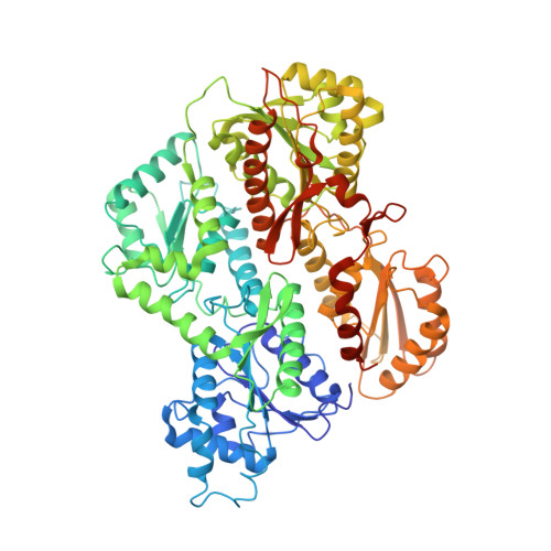
    4OMT

  • PubMed Abstract: 

    Whereas the three-dimensional structure and the structural basis of the allosteric regulation of prokaryotic 6-phosphofructokinases (Pfks) have been studied in great detail, knowledge of the molecular basis of the allosteric behaviour of the far more complex mammalian Pfks is still very limited. The human muscle isozyme was expressed heterologously in yeast cells and purified using a five-step purification protocol. Protein crystals suitable for diffraction experiments were obtained by the vapour-diffusion method. The crystals belonged to space group P6222 and diffracted to 6.0 Å resolution. The 3.2 Å resolution structure of rabbit muscle Pfk (rmPfk) was placed into the asymmetric unit and optimized by rigid-body and group B-factor refinement. Interestingly, the tetrameric enzyme dissociated into a dimer, similar to the situation observed in the structure of rmPfk.


  • Organizational Affiliation
    • Institute of Bioanalytical Chemistry, Center for Biotechnology and Biomedicine, University of Leipzig, Deutscher Platz 5, 04103 Leipzig, Germany.

Macromolecule Content 

  • Total Structure Weight: 85.29 kDa 
  • Atom Count: 5,715 
  • Modeled Residue Count: 748 
  • Deposited Residue Count: 780 
  • Unique protein chains: 1

Macromolecules

Find similar proteins by:|  3D Structure
Entity ID: 1
MoleculeChains  Sequence LengthOrganismDetailsImage
6-phosphofructokinase, muscle type780Homo sapiensMutation(s): 0 
Gene Names: PFKMPFKX
EC: 2.7.1.11
UniProt & NIH Common Fund Data Resources
Find proteins for P08237 (Homo sapiens)
Explore P08237 
Go to UniProtKB:  P08237
PHAROS:  P08237
GTEx:  ENSG00000152556 
Entity Groups
Sequence Clusters30% Identity50% Identity70% Identity90% Identity95% Identity100% Identity
UniProt GroupP08237
Sequence Annotations
Expand
Reference Sequence

Experimental Data & Validation

Experimental Data

  • Method: X-RAY DIFFRACTION
  • Resolution: 6.00 Å
  • R-Value Free:  0.274 (Depositor), 0.291 (DCC) 
  • R-Value Work:  0.238 (Depositor), 0.263 (DCC) 
  • R-Value Observed: 0.242 (Depositor) 
Space Group: P 62 2 2
Unit Cell:
Length ( Å )Angle ( ˚ )
a = 229.7α = 90
b = 229.7β = 90
c = 133γ = 120
Software Package:
Software NamePurpose
PHENIXrefinement
PHASERphasing
REFMACrefinement
XDSdata reduction
Aimlessdata scaling
MxCuBEdata collection

Structure Validation

View Full Validation Report


Entry History 

Deposition Data

Revision History  (Full details and data files)

  • Version 1.0: 2014-05-14
    Type: Initial release
  • Version 1.1: 2014-09-24
    Changes: Database references
  • Version 1.2: 2014-12-10
    Changes: Structure summary
  • Version 1.3: 2019-07-17
    Changes: Data collection, Refinement description
  • Version 1.4: 2023-09-20
    Changes: Data collection, Database references, Refinement description