4MXP | pdb_00004mxp

Structural Basis for PI(4)P-Specific Membrane Recruitment of the Legionella pneumophila Effector DrrA/SidM


Experimental Data Snapshot

  • Method: X-RAY DIFFRACTION
  • Resolution: 1.83 Å
  • R-Value Free: 
    0.208 (Depositor), 0.221 (DCC) 
  • R-Value Work: 
    0.189 (Depositor), 0.198 (DCC) 
  • R-Value Observed: 
    0.190 (Depositor) 

Starting Model: experimental
View more details

wwPDB Validation 3D Report Full Report

Validation slider image for 4MXP

Ligand Structure Quality Assessment 


This is version 1.2 of the entry. See complete history

Literature

Structural Basis for PI(4)P-Specific Membrane Recruitment of the Legionella pneumophila Effector DrrA/SidM.

Del Campo, C.M.Mishra, A.K.Wang, Y.H.Roy, C.R.Janmey, P.A.Lambright, D.G.

(2014) Structure 22: 397-408

  • DOI: https://doi.org/10.1016/j.str.2013.12.018
  • Primary Citation Related Structures: 
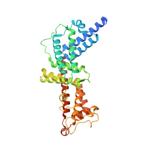
    4MXP

  • PubMed Abstract: 

    Recruitment of the Legionella pneumophila effector DrrA to the Legionella-containing vacuole, where it activates and AMPylates Rab1, is mediated by a P4M domain that binds phosphatidylinositol 4-phosphate [PI(4)P] with high affinity and specificity. Despite the importance of PI(4)P in Golgi trafficking and its manipulation by pathogens, the structural bases for PI(4)P-dependent membrane recruitment remain poorly defined. Here, we determined the crystal structure of a DrrA fragment including the P4M domain in complex with dibutyl PI(4)P and investigated the determinants of phosphoinositide recognition and membrane targeting. Headgroup recognition involves an elaborate network of direct and water-mediated interactions with basic and polar residues in the context of a deep, constrictive binding pocket. An adjacent hydrophobic helical element packs against the acyl chains and inserts robustly into PI(4)P-containing monolayers. The structural, biochemical, and biophysical data reported here support a detailed structural mechanism for PI(4)P-dependent membrane targeting by DrrA.


  • Organizational Affiliation
    • Program in Molecular Medicine and Department of Biochemistry and Molecular Pharmacology, University of Massachusetts Medical School, Worcester, MA 01605, USA.

Macromolecule Content 

  • Total Structure Weight: 37.69 kDa 
  • Atom Count: 2,696 
  • Modeled Residue Count: 310 
  • Deposited Residue Count: 328 
  • Unique protein chains: 1

Macromolecules

Find similar proteins by:|  3D Structure
Entity ID: 1
MoleculeChains  Sequence LengthOrganismDetailsImage
Defects in Rab1 recruitment protein A328Legionella pneumophila subsp. pneumophila str. Philadelphia 1Mutation(s): 0 
Gene Names: drrAlpg2464sidM
EC: 2.7.7 (PDB Primary Data), 2.7.7.108 (UniProt)
Membrane Entity: Yes 
UniProt
Find proteins for Q5ZSQ3 (Legionella pneumophila subsp. pneumophila (strain Philadelphia 1 / ATCC 33152 / DSM 7513))
Explore Q5ZSQ3 
Go to UniProtKB:  Q5ZSQ3
Entity Groups
Sequence Clusters30% Identity50% Identity70% Identity90% Identity95% Identity100% Identity
UniProt GroupQ5ZSQ3
Sequence Annotations
Expand
Reference Sequence

Small Molecules

Ligands 2 Unique
IDChains Name / Formula / InChI Key2D Diagram3D Interactions
DB4

Query on DB4


Download:Ideal Coordinates CCD File
B [auth A](2R)-3-{[(R)-hydroxy{[(1R,2R,3R,4R,5S,6R)-2,3,5,6-tetrahydroxy-4-(phosphonooxy)cyclohexyl]oxy}phosphoryl]oxy}propane-1,2-diyl dibutanoate
C17 H32 O16 P2
GZEWZLHSHLUSGP-HDIZFQRBSA-N
NA

Query on NA


Download:Ideal Coordinates CCD File
C [auth A]SODIUM ION
Na
FKNQFGJONOIPTF-UHFFFAOYSA-N

Experimental Data & Validation

Experimental Data

  • Method: X-RAY DIFFRACTION
  • Resolution: 1.83 Å
  • R-Value Free:  0.208 (Depositor), 0.221 (DCC) 
  • R-Value Work:  0.189 (Depositor), 0.198 (DCC) 
  • R-Value Observed: 0.190 (Depositor) 
Space Group: I 41
Unit Cell:
Length ( Å )Angle ( ˚ )
a = 128.552α = 90
b = 128.552β = 90
c = 53.222γ = 90
Software Package:
Software NamePurpose
REFMACrefinement
PDB_EXTRACTdata extraction
HKL-2000data collection
DENZOdata reduction
SCALEPACKdata scaling
PHENIXphasing

Structure Validation

View Full Validation Report


Ligand Structure Quality Assessment 


Entry History 

Deposition Data

Revision History  (Full details and data files)

  • Version 1.0: 2014-03-19
    Type: Initial release
  • Version 1.1: 2014-03-26
    Changes: Database references
  • Version 1.2: 2023-09-20
    Changes: Data collection, Database references, Derived calculations, Refinement description