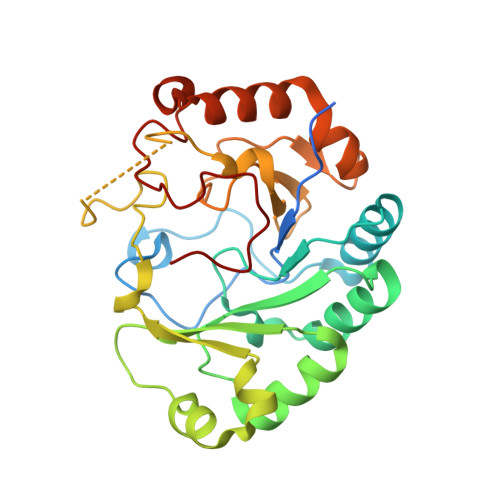
4M7T

Crystal structure of BtrN in complex with AdoMet and 2-DOIA


Experimental Data Snapshot

  • Method: X-RAY DIFFRACTION
  • Resolution: 1.56 Å
  • R-Value Free: 0.182 
  • R-Value Work: 0.164 
  • R-Value Observed: 0.165 

wwPDB Validation   3D Report Full Report


Ligand Structure Quality Assessment 


This is version 1.3 of the entry. See complete history


Literature

X-ray analysis of butirosin biosynthetic enzyme BtrN redefines structural motifs for AdoMet radical chemistry.

Goldman, P.J.Grove, T.L.Booker, S.J.Drennan, C.L.

(2013) Proc Natl Acad Sci U S A 110: 15949-15954

  • DOI: https://doi.org/10.1073/pnas.1312228110
  • Primary Citation of Related Structures:  
    4M7S, 4M7T

  • PubMed Abstract: 

    The 2-deoxy-scyllo-inosamine (DOIA) dehydrogenases are key enzymes in the biosynthesis of 2-deoxystreptamine-containing aminoglycoside antibiotics. In contrast to most DOIA dehydrogenases, which are NAD-dependent, the DOIA dehydrogenase from Bacillus circulans (BtrN) is an S-adenosyl-l-methionine (AdoMet) radical enzyme. To examine how BtrN employs AdoMet radical chemistry, we have determined its structure with AdoMet and substrate to 1.56 Å resolution. We find a previously undescribed modification to the core AdoMet radical fold: instead of the canonical (β/α)6 architecture, BtrN displays a (β5/α4) motif. We further find that an auxiliary [4Fe-4S] cluster in BtrN, thought to bind substrate, is instead implicated in substrate-radical oxidation. High structural homology in the auxiliary cluster binding region between BtrN, fellow AdoMet radical dehydrogenase anSME, and molybdenum cofactor biosynthetic enzyme MoaA provides support for the establishment of an AdoMet radical structural motif that is likely common to ~6,400 uncharacterized AdoMet radical enzymes.


  • Organizational Affiliation

    Departments of Chemistry and Biology and Howard Hughes Medical Institute, Massachusetts Institute of Technology, Cambridge, MA 02139.


Macromolecules
Find similar proteins by:  (by identity cutoff)  |  3D Structure
Entity ID: 1
MoleculeChains Sequence LengthOrganismDetailsImage
BtrN270Niallia circulansMutation(s): 0 
Gene Names: btrN
EC: 1.1.99.38
UniProt
Find proteins for Q8G907 (Niallia circulans)
Explore Q8G907 
Go to UniProtKB:  Q8G907
Entity Groups  
Sequence Clusters30% Identity50% Identity70% Identity90% Identity95% Identity100% Identity
UniProt GroupQ8G907
Sequence Annotations
Expand
  • Reference Sequence
Small Molecules
Ligands 4 Unique
IDChains Name / Formula / InChI Key2D Diagram3D Interactions
SAM
Query on SAM

Download Ideal Coordinates CCD File 
D [auth A]S-ADENOSYLMETHIONINE
C15 H22 N6 O5 S
MEFKEPWMEQBLKI-FCKMPRQPSA-N
SF4
Query on SF4

Download Ideal Coordinates CCD File 
B [auth A],
C [auth A]
IRON/SULFUR CLUSTER
Fe4 S4
LJBDFODJNLIPKO-UHFFFAOYSA-N
25W
Query on 25W

Download Ideal Coordinates CCD File 
E [auth A](1R,2S,3S,4R,5S)-5-aminocyclohexane-1,2,3,4-tetrol
C6 H13 N O4
QXQNRSUOYNMXDL-KGJVWPDLSA-N
GOL
Query on GOL

Download Ideal Coordinates CCD File 
F [auth A]GLYCEROL
C3 H8 O3
PEDCQBHIVMGVHV-UHFFFAOYSA-N
Experimental Data & Validation

Experimental Data

  • Method: X-RAY DIFFRACTION
  • Resolution: 1.56 Å
  • R-Value Free: 0.182 
  • R-Value Work: 0.164 
  • R-Value Observed: 0.165 
  • Space Group: P 21 21 21
Unit Cell:
Length ( Å )Angle ( ˚ )
a = 44.664α = 90
b = 55.175β = 90
c = 116.853γ = 90
Software Package:
Software NamePurpose
DENZOdata reduction
SCALEPACKdata scaling
PHASERphasing
PHENIXrefinement
PDB_EXTRACTdata extraction
APEXdata collection
HKL-2000data reduction
HKL-2000data scaling

Structure Validation

View Full Validation Report


Ligand Structure Quality Assessment 


Entry History 

Deposition Data

Revision History  (Full details and data files)

  • Version 1.0: 2013-10-02
    Type: Initial release
  • Version 1.1: 2014-02-05
    Changes: Database references
  • Version 1.2: 2017-11-15
    Changes: Refinement description
  • Version 1.3: 2024-02-28
    Changes: Data collection, Database references, Derived calculations